. 2017 Jan;14(1):17-25.
doi: 10.1513/AnnalsATS.201606-459OC.

Chest Computed Tomographic Image Screening for Cystic Lung Diseases in Patients with Spontaneous Pneumothorax Is Cost Effective

Affiliations

Chest Computed Tomographic Image Screening for Cystic Lung Diseases in Patients with Spontaneous Pneumothorax Is Cost Effective

Nishant Gupta et al. Ann Am Thorac Soc. 2017 Jan.

Abstract

Rationale: Patients without a known history of lung disease presenting with a spontaneous pneumothorax are generally diagnosed as having primary spontaneous pneumothorax. However, occult diffuse cystic lung diseases such as Birt-Hogg-Dubé syndrome (BHD), lymphangioleiomyomatosis (LAM), and pulmonary Langerhans cell histiocytosis (PLCH) can also first present with a spontaneous pneumothorax, and their early identification by high-resolution computed tomographic (HRCT) chest imaging has implications for subsequent management.

Objectives: The objective of our study was to evaluate the cost-effectiveness of HRCT chest imaging to facilitate early diagnosis of LAM, BHD, and PLCH.

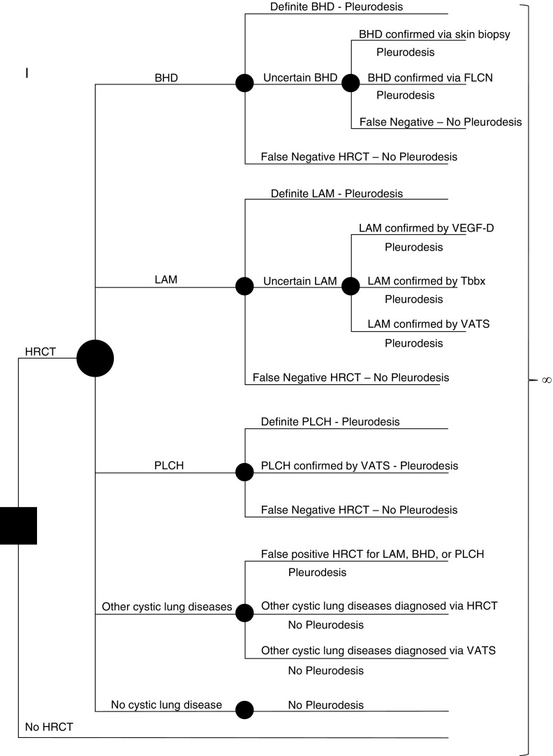
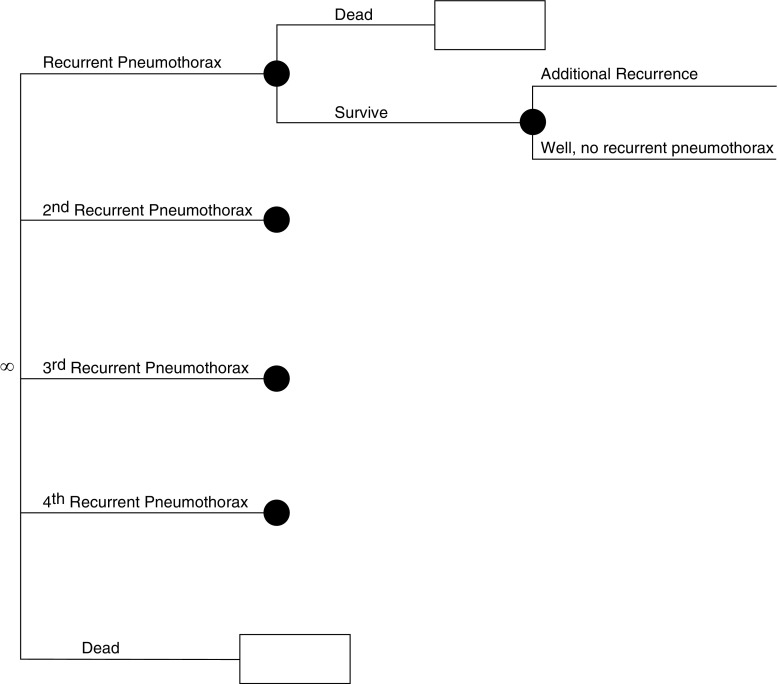
Methods: We constructed a Markov state-transition model to assess the cost-effectiveness of screening HRCT to facilitate early diagnosis of diffuse cystic lung diseases in patients presenting with an apparent primary spontaneous pneumothorax. Baseline data for prevalence of BHD, LAM, and PLCH and rates of recurrent pneumothoraces in each of these diseases were derived from the literature. Costs were extracted from 2014 Medicare data. We compared a strategy of HRCT screening followed by pleurodesis in patients with LAM, BHD, or PLCH versus conventional management with no HRCT screening.

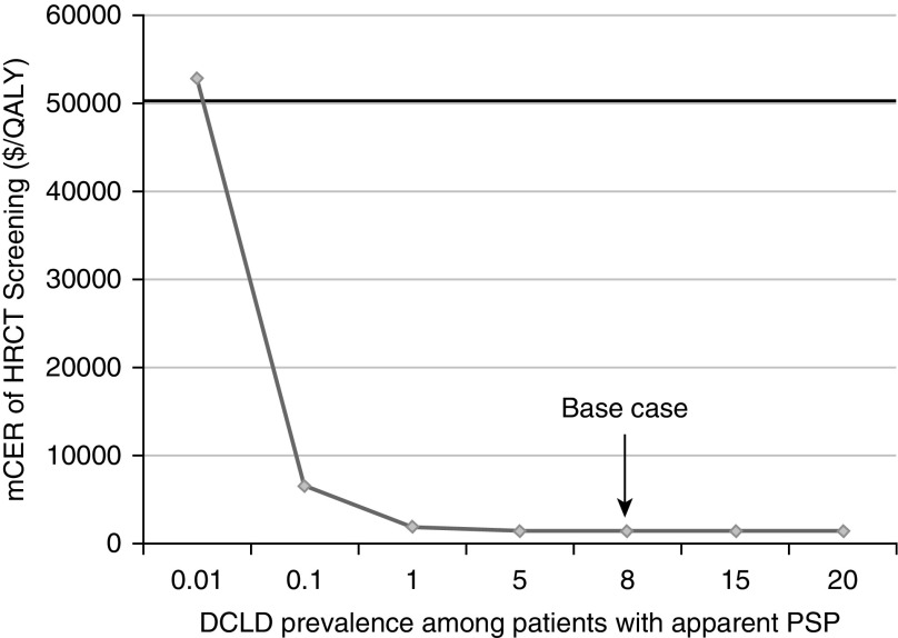
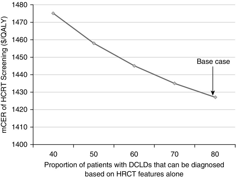
Measurements and main results: In our base case analysis, screening for the presence of BHD, LAM, or PLCH in patients presenting with a spontaneous pneumothorax was cost effective, with a marginal cost-effectiveness ratio of $1,427 per quality-adjusted life-year gained. Sensitivity analysis showed that screening HRCT remained cost effective for diffuse cystic lung diseases prevalence as low as 0.01%.

Conclusions: HRCT image screening for BHD, LAM, and PLCH in patients with apparent primary spontaneous pneumothorax is cost effective. Clinicians should consider performing a screening HRCT in patients presenting with apparent primary spontaneous pneumothorax.

Keywords: Birt-Hogg-Dubé syndrome; lymphangioleiomyomatosis; pneumothorax; pulmonary Langerhans cell histiocytosis.


Figures

Figure 1.
Figure 1.
Simplified schematic of the model used in our analysis. The solid square represents the decision node where the initial decision is made by the practicing clinician—to obtain a high-resolution computed tomography (HRCT) scan or not. Each solid circle represents a chance node, where the likelihood of a given outcome is defined by a probability entered into the model. Patients presenting with a pneumothorax can have one of the following five scenarios: underlying Birt-Hogg-Dubé syndrome (BHD), underlying lymphangioleiomyomatosis (LAM), underlying pulmonary Langerhans cell histiocytosis (PLCH), other cystic lung diseases, or no cystic lung disease. The probability of the presence of each of these five scenarios is entered into the model. Further testing for diagnostic confirmation is based on the sensitivity and specificity of HRCT to accurately diagnose LAM, BHD, or PLCH. The performance characteristics of each diagnostic test are also considered when assigning a correct diagnosis, and the rates of false-positive and false-negative diagnoses are accounted for at each step. Each arm of the tree ultimately ends in the Markov node (∞ symbol). In each state, costs and quality adjustment factors are applied and tabulated. FLCN = folliculin; Tbbx = transbronchial biopsy; VATS = video-assisted thoracoscopic surgery; VEGF-D = vascular endothelial growth factor-D.
Figure 2.
Figure 2.
Simplified schematic of the model used in our analysis. This figure depicts the continuation of our model. In the Markov node (∞ symbol), patients progress through various health states dictated by probabilities entered into the model. These probabilities are different, depending on how the individual entered the Markov node (e.g., a patient with cystic lung disease [lymphangioleiomyomatosis (LAM), Birt-Hogg-Dubé syndrome (BHD), or pulmonary Langerhans cell histiocytosis (PLCH)] who has undergone pleurodesis has a lower probability of recurrent pneumothorax than a patient who has not). In each cycle (defined as 1 mo), patients are subjected to risk of recurrent pneumothorax or age/sex-related death, as well as disease-specific additional mortality as applicable to patients with LAM and PLCH. In each state, costs and quality adjustment factors are applied and tabulated. These cycles repeat until all patients are dead. Outcomes can then be compared for each strategy tested (high-resolution computed tomography [HRCT] vs. no HRCT).
Figure 3.
Figure 3.
Marginal cost-effectiveness ratio (mCER) of high-resolution computed tomography (HRCT) chest screening in relationship to the prevalence of diffuse cystic lung diseases (DCLDs) in patients presenting with an apparent primary spontaneous pneumothorax (PSP). This analysis demonstrates that as the prevalence of DCLDs among patients with an apparent PSP increases, it becomes more cost effective to screen for the presence of DCLDs. By convention, treatment strategies with an mCER of less than $50,000 per quality-adjusted life-year (QALY) gained are considered cost effective. On the basis of that threshold, the strategy for HRCT screening remains cost effective as long as DCLD prevalence among patients presenting with an apparent PSP is greater than 0.01%. Arrow denotes the base case in our analysis.
Figure 4.
Figure 4.
Marginal cost-effectiveness ratio (mCER) of high-resolution computed tomography (HRCT) chest screening in relationship to the ability to accurately diagnose the exact diffuse cystic lung disease (DCLD) on the basis of imaging features alone. This analysis demonstrates that as the sensitivity of HRCT to diagnose DCLD increases, the strategy of HRCT screening becomes more cost effective. However, at all sensitivity values of HRCT tested in our analysis, the strategy of HRCT screening remained cost effective, with an mCER substantially below the conventional cost-effectiveness threshold of $50,000 per quality-adjusted life-year (QALY) gained. Arrow denotes the base case in our analysis.

Comment in

References

    1. Gupta N, Vassallo R, Wikenheiser-Brokamp KA, McCormack FX. Diffuse cystic lung disease: part I. Am J Respir Crit Care Med. 2015;191:1354–1366. - PMC - PubMed
    1. Gupta D, Hansell A, Nichols T, Duong T, Ayres JG, Strachan D. Epidemiology of pneumothorax in England. Thorax. 2000;55:666–671. - PMC - PubMed
    1. Melton LJ, III, Hepper NG, Offord KP. Incidence of spontaneous pneumothorax in Olmsted County, Minnesota: 1950 to 1974. Am Rev Respir Dis. 1979;120:1379–1382. - PubMed
    1. Baumann MH, Strange C, Heffner JE, Light R, Kirby TJ, Klein J, Luketich JD, Panacek EA, Sahn SA, Group APC AACP Pneumothorax Consensus Group. Management of spontaneous pneumothorax: an American College of Chest Physicians Delphi consensus statement. Chest. 2001;119:590–602. - PubMed
    1. Bintcliffe OJ, Hallifax RJ, Edey A, Feller-Kopman D, Lee YC, Marquette CH, Tschopp JM, West D, Rahman NM, Maskell NA. Spontaneous pneumothorax: time to rethink management? Lancet Respir Med. 2015;3:578–588. - PubMed

MeSH terms