

PD-L1 is upregulated by EBV-driven LMP1 through NF-κB pathway and correlates with poor prognosis in natural killer/T-cell lymphoma
- PMID: 27737703
- PMCID: PMC5064887
- DOI: 10.1186/s13045-016-0341-7
PD-L1 is upregulated by EBV-driven LMP1 through NF-κB pathway and correlates with poor prognosis in natural killer/T-cell lymphoma
Abstract
Background: Natural killer/T-cell lymphoma (NKTCL) is an Epstein-Barr virus (EBV)-associated, highly aggressive lymphoma. Treatment outcome remains sub-optimal, especially for advanced-stage or relapsed diseases. Programmed cell death receptor 1 (PD-1) and PD ligand 1 (PD-L1) have become promising therapeutic targets for various malignancies, but their role in the pathogenesis and their interactions with EBV in NKTCL remains to be investigated.
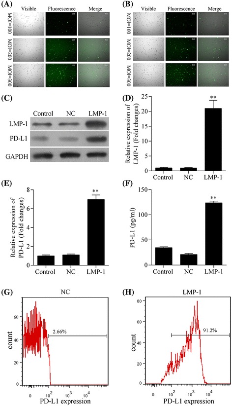
Methods: Expression of PD-L1 was measured in NK-92 (EBV-negative) and SNK-6 (EBV-positive) cells by western blot, quantitative real-time PCR and enzyme-linked immunosorbent assay, and flow cytometry, respectively. Latent membrane protein 1 (LMP1)-harboring lentiviral vectors were transfected into NK-92 cells to examine the correlation between LMP1 and PD-L1 expression. Proteins in the downstream pathways of LMP1 signaling were measured in NK-92 cells transfected with LMP1-harboring or negative control vectors as well as in SNK-6 cells. PD-L1 expression on tumor specimens and serum concentration of soluble PD-L1 were collected in a retrospective cohort of patients with Ann Arbor stage I~II NKTCL, and their prognostic significance were analyzed.
Results: Expression of PD-L1 was significantly higher in SNK-6 cells than in NK-92 cells, at both protein and mRNA levels. Expression of PD-L1 was remarkably upregulated in NK-92 cells transfected with LMP1-harboring lentiviral vectors compared with those transfected with negative control vectors. Proteins in the MAPK/NF-κB pathway were upregulated in LMP1-expressing NK-92 cells compared with the negative control. Selective inhibitors of those proteins induced significant downregulation of PD-L1 expression in LMP1-expressing NK-92 cells as well as in SNK-6 cells. Patients with a high concentration of serum soluble PD-L1 (≥3.4 ng/ml) or with a high percentage of PD-L1 expression in tumor specimens (≥38 %) exhibited significantly lower response rate to treatment and remarkably worse survival, compared with their counterparts. A high concentration of serum soluble PD-L1 and a high percentage of PD-L1 expression in tumor specimens were independent adverse prognostic factors among patients with stage I~II NKTCL.
Conclusions: PD-L1 expression positively correlated LMP1 expression in NKTCL, which was probably mediated by the MAPK/NF-κB pathway. PD-L1 expression in serum and tumor tissues has significant prognostic value for early-stage NKTCL.
Keywords: Epstein–Barr virus; Latent membrane protein 1; Natural killer/T-cell lymphoma; Programmed cell death receptor 1.
Figures


References
-
- Chan JK, Quintanilla-Martinez L, Ferry JA, Peh S-C, et al. Extranodal NK/T-cell lymphoma, nasal type. In: Swerdlow SH, Campo E, Harris NL, et al., editors. WHO classification of tumours of haematopoietic and lymphoid tissues. Lyon: IARC; 2008. pp. 285–288.
-
- Chan JK, Jaffe ES, Ralfkiaer E. Extranodal NK/T-cell lymphoma, nasal type. In: Jaffe ES, Harris NL, Stein H, Vardiman JW, editors. WHO classification of tumours of haematopoietic and lymphoid tissues. Lyon: IARC; 2001. pp. 204–207.
-
- Au WY, Weisenburger DD, Intragumtornchai T, Nakamura S, Kim WS, Sng I, et al. Clinical differences between nasal and extranasal natural killer/T-cell lymphoma: a study of 136 cases from the International Peripheral T-Cell Lymphoma Project. Blood. 2009;113:3931–3937. doi: 10.1182/blood-2008-10-185256. - DOI - PubMed
-
- Bi XW, Li YX, Fang H, Jin J, Wang WH, Wang SL, et al. High-dose and extended-field intensity modulated radiation therapy for early-stage NK/T-cell lymphoma of Waldeyer’s ring: dosimetric analysis and clinical outcome. Int J Radiat Oncol Biol Phys. 2013;87:1086–1093. doi: 10.1016/j.ijrobp.2013.08.040. - DOI - PubMed
Publication types
MeSH terms
Substances
LinkOut - more resources
Full Text Sources
Other Literature Sources
Research Materials
