. 2017 Mar 13;26(3):417-427.
doi: 10.3727/096368916X693464. Epub 2016 Oct 12.

Chondrogenic Differentiation of Mesenchymal Stem Cells in Three-Dimensional Chitosan Film Culture

Chondrogenic Differentiation of Mesenchymal Stem Cells in Three-Dimensional Chitosan Film Culture

Tsai-Jung Lu et al. Cell Transplant. .

Abstract

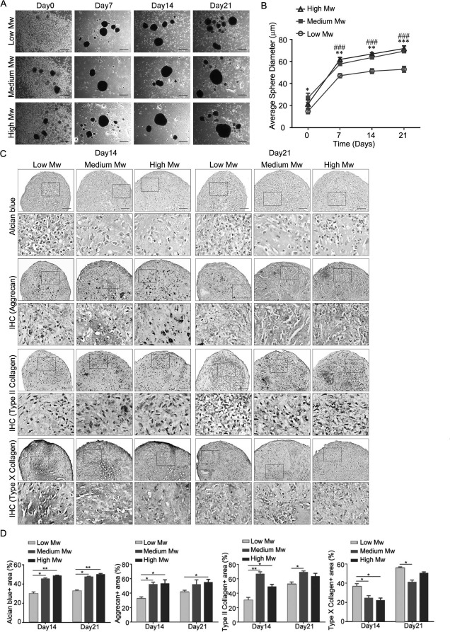
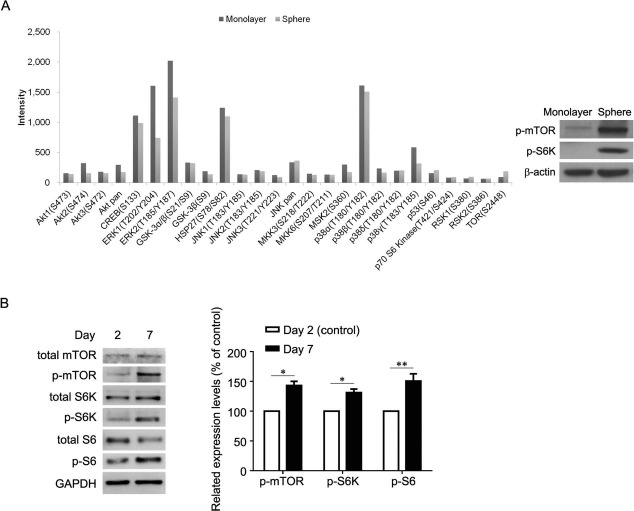
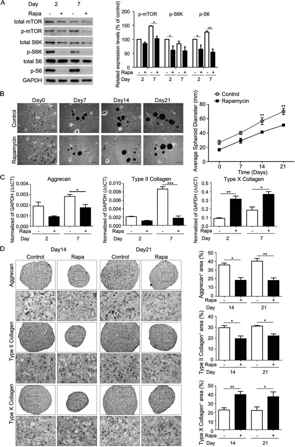
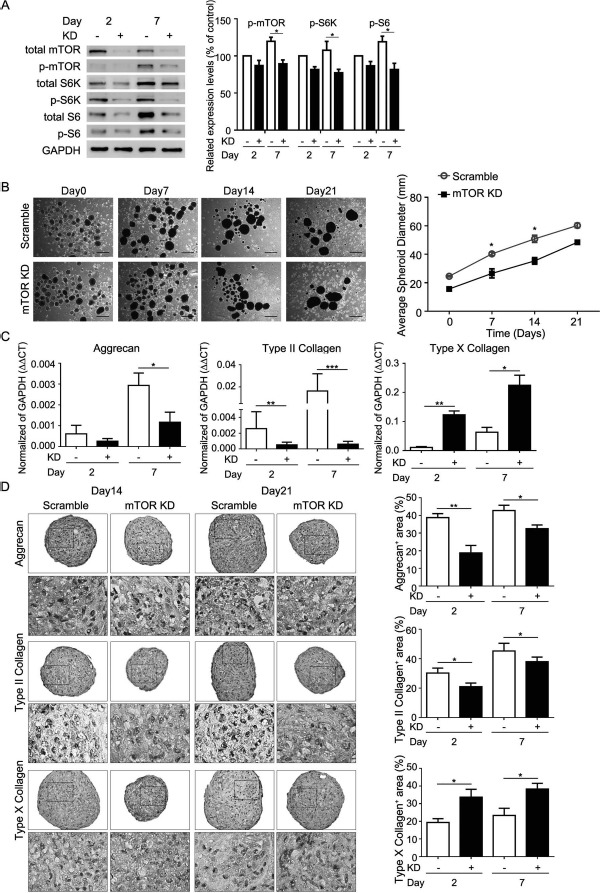
Articular cartilage has a very limited capacity for self-repair, and mesenchymal stem cells (MSCs) have the potential to treat cartilage defects and osteoarthritis. However, in-depth mechanistic studies regarding their applications are required. Here we demonstrated the use of chitosan film culture for promoting chondrogenic differentiation of MSCs. We found that MSCs formed spheres 2 days after seeding on dishes coated with chitosan. When MSCs were induced in a chondrogenic induction medium on chitosan films, the size of the spheres continuously increased for up to 21 days. Alcian blue staining and immunohistochemistry demonstrated the expression of chondrogenic proteins, including aggrecan, type II collagen, and type X collagen at 14 and 21 days of differentiation. Importantly, chitosan, with a medium molecular weight (size: 190-310 kDa), was more suitable than other sizes for inducing chondrogenic differentiation of MSCs in terms of sphere size and expression of chondrogenic proteins and endochondral markers. We identified that the mechanistic target of rapamycin (mTOR) signaling and its downstream S6 kinase (S6K)/S6 were activated in chitosan film culture compared to that of monolayer culture. The activation of mTOR/S6K was continuously upregulated from days 2 to 7 of differentiation. Furthermore, we found that mTOR/S6K signaling was required for chondrogenic differentiation of MSCs in chitosan film culture through rapamycin treatment and mTOR knockdown. In conclusion, we showed the suitability of chitosan film culture for promoting chondrogenic differentiation of MSCs and its potential in the development of new strategies in cartilage tissue engineering.


Figures

Figure 1.
Figure 1.
Effects of different molecular weight chitosan films on morphology and chondrogenic protein expression in MSC spheroid. Aliquots of 2× 10⁵ mesenchymal stem cells (MSCs) in complete culture medium were seeded to each well of a six-well plate, coated with different molecular weight (Mw) chitosan. Medium was replaced with chondrogenic induction medium 2 days later (Day0). (A) Morphology, (B) average spheroid diameter, and (C) Alcian blue or immunohistochemistry of indicated proteins in MSC spheroids on different Mw chitosan films for indicated time of differentiation. (D) Quantification of the data in (C). Quantification data are presented as mean+standard error of the mean (SEM) (n = 3). p < 0.05, ∗∗p < 0.01, ∗∗∗p < 0.001, ###p < 0.001 (medium Mw vs. low Mw) as determined by one-way analysis of variance (ANOVA). Scale bar: 500 μm.
Figure 2.
Figure 2.
Signaling differences between monolayer and spheroid culture. (A, left) Human phospho-MAPK array and (A, right) Western blot analysis for the signaling differences between monolayer and spheroid culture. (B, left) Western blot analysis and (B, right) its quantification for indicated periods of chondrogenesis. p < 0.05, ∗∗p < 0.01 as determined by paired Student's t-test. Monolayer, monolayer culture; Sphere, spheroid culture; Day 2, chondrogenic induction medium cultured for 2 days; Day 7, chondrogenic induction medium cultured for 7 days.
Figure 3.
Figure 3.
Effects of rapamycin on size and chondrogenic protein expression of MSC spheroid on chitosan films. Aliquots of 2× 10⁵ MSCs in complete culture medium were seeded to each well of a six-well plate, coated with chitosan. Medium was replaced with chondrogenic induction medium in the absence or presence of rapamycin (Rapa) 2 days later (Day0). (A, left) Western blot analysis of mTOR-related signaling proteins and (A, right) its quantification at indicated time periods. p < 0.05, ∗∗p < 0.01 as determined by paired Student's t-test. (B, left) Morphology, (B, right) average spheroid diameter, and (C) quantitative RT-PCR for mRNA levels. p < 0.05, ∗∗p < 0.01, ∗∗∗p < 0.001 as determined by paired Student's t-test. (D, left) Immunohistochemistry for indicated proteins of MSC spheroids at indicated time periods. (D, right) Quantification data of immunohistochemistry. Data are presented as mean ± SEM (n = 3). p < 0.05, ∗∗p < 0.01, ∗∗∗p < 0.001 as determined by unpaired Student's t-test. Scale bar: 500 μm. Day2, chondrogenic induction medium cultured for 2 days; Day7, chondrogenic induction medium cultured for 7 days; Day 14, chondrogenic induction medium cultured for 14 days; Day21, chondrogenic induction medium cultured for 21 days; Rapa, chondrogenic induction medium in the presence of rapamycin; Control, chondrogenic induction medium in the absence of rapamycin.
Figure 4.
Figure 4.
Effects of mTOR knockdown (KD) on size and chondrogenic protein expression of MSC spheroid on chitosan films. Aliquots of 2 × 10⁵ MSCs with or without mTOR KD in complete culture medium were seeded to each well of a six-well plate, coated with chitosan. Medium was replaced with chondrogenic induction medium 2 days later (Day0). (A, left) Western blot analysis of mTOR-related signaling proteins and (A, right) its quantification at indicated time periods. p < 0.05 as determined by paired Student's t-test. (B, left) Morphology, (B, right) average spheroid diameter, (C) quantitative RT-PCR for mRNA levels, and (D, left) immunohistochemistry for indicated proteins of MSC spheroids at indicated time periods. (D, right) Quantification data of immunohistochemistry. Data are presented as mean + SEM (n = 3). p < 0.05, ∗∗p < 0.01, ∗∗∗p < 0.001 as determined by unpaired Student's t-test. Scale bar: 500 μm. Day2, chondrogenic induction medium cultured for 2 days; Day7, chondrogenic induction medium cultured for 7 days; Day14, chondrogenic induction medium cultured for 14 days; Day21, chondrogenic induction medium cultured for 21 days; KD, MSCs with mTOR KD; Scramble, MSCs without mTOR KD.

References

    1. Wakitani S, Goto T, Young RG, Mansour JM, Goldberg VM, Caplan AI. Repair of large full-thickness articular cartilage defects with allograft articular chondrocytes embedded in a collagen gel. Tissue Eng. 1998; 4: 429–44. - PubMed
    1. Brittberg M, Lindahl A, Nilsson A, Ohlsson C, Isaksson O, Peterson L. Treatment of deep cartilage defects in the knee with autologous chondrocyte transplantation. N Engl J Med. 1994; 331: 889–95. - PubMed
    1. Prockop DJ. Marrow stromal cells as stem cells for non-hematopoietic tissues. Science 1997; 276: 71–4. - PubMed
    1. Hung SC, Cheng H, Pan CY, Tsai MJ, Kao LS, Ma HL. In vitro differentiation of size-sieved stem cells into electrically active neural cells. Stem Cells 2002; 20: 522–9. - PubMed
    1. Caplan AI. Review: Mesenchymal stem cells: Cell-based reconstructive therapy in orthopedics. Tissue Eng. 2005; 11: 1198–211. - PubMed

MeSH terms

LinkOut - more resources