

Cognitive assessment of pycnogenol therapy following traumatic brain injury
- PMID: 27737807
- PMCID: PMC5131724
- DOI: 10.1016/j.neulet.2016.10.016
Cognitive assessment of pycnogenol therapy following traumatic brain injury
Abstract
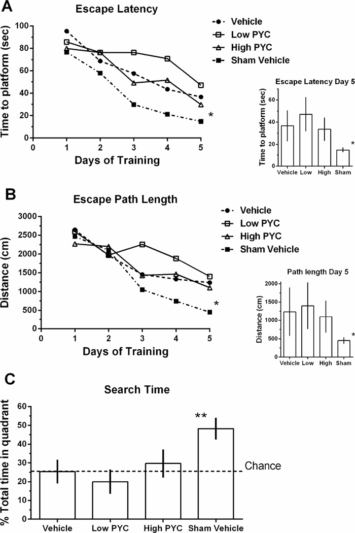
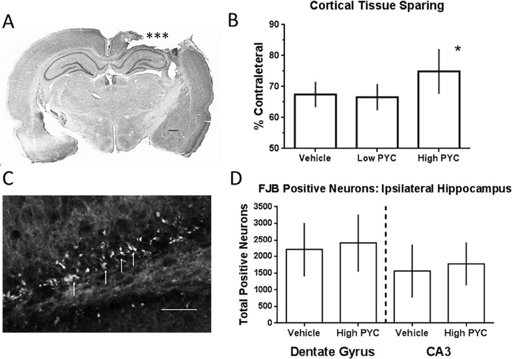
We have previously shown that pycnogenol (PYC) increases antioxidants, decreases oxidative stress, suppresses neuroinflammation and enhances synaptic plasticity following traumatic brain injury (TBI). Here, we investigate the effects of PYC on cognitive function following a controlled cortical impact (CCI). Adult Sprague-Dawley rats received a CCI injury followed by an intraperitoneal injection of PYC (50 or 100mg/kg). Seven days post trauma, subjects were evaluated in a Morris water maze (MWM) and evaluated for changes in lesion volume. Some animals were evaluated at 48h for hippocampal Fluoro-jade B (FJB) staining. The highest dose of PYC therapy significantly reduced lesion volume, with no improvement in MWM compared to vehicle controls. PYC failed to reduce the total number of FJB positive neurons in the hippocampus. These results suggest that the reduction of oxidative stress and neuroinflammation are not the key components of the secondary injury that contribute to cognitive deficits following TBI.
Keywords: Bioflavonoids; Cortical contusion; Head injury; Natural compounds; Recovery of function; Water maze.
Copyright © 2016 Elsevier Ireland Ltd. All rights reserved.
Conflict of interest statement
All authors declare no conflict of interest.
Figures
References
-
- Alder J, Fujioka W, Giarratana A, Wissocki J, Thakkar K, Vuong P, Patel B, Chakraborty T, Elsabeh R, Parikh A, Girn HS, Crockett D, Thakker-Varia S. Genetic and pharmacological intervention of the p75NTR pathway alters morphological and behavioural recovery following traumatic brain injury in mice. Brain Inj. 2016;30:48–65. - PubMed
-
- Amaral D, Witter M. Hippocampal formation. In: Paxinos G, editor. The rat nervous system. New York: Academic Press; 1995. pp. 443–493.
-
- Anderson KJ, Miller KM, Fugaccia I, Scheff SW. Regional distribution of fluoro-jade B staining in the hippocampus following traumatic brain injury. Exp. Neurol. 2005;193:125–130. - PubMed
MeSH terms
Substances
Grants and funding
LinkOut - more resources
Full Text Sources
Other Literature Sources
Medical
