. 2016 Nov 15;134(20):1557-1567.
doi: 10.1161/CIRCULATIONAHA.114.014998. Epub 2016 Oct 13.

Mechanical Stress Conditioning and Electrical Stimulation Promote Contractility and Force Maturation of Induced Pluripotent Stem Cell-Derived Human Cardiac Tissue

Affiliations

Mechanical Stress Conditioning and Electrical Stimulation Promote Contractility and Force Maturation of Induced Pluripotent Stem Cell-Derived Human Cardiac Tissue

Jia-Ling Ruan et al. Circulation. .

Abstract

Background: Tissue engineering enables the generation of functional human cardiac tissue with cells derived in vitro in combination with biocompatible materials. Human-induced pluripotent stem cell-derived cardiomyocytes provide a cell source for cardiac tissue engineering; however, their immaturity limits their potential applications. Here we sought to study the effect of mechanical conditioning and electric pacing on the maturation of human-induced pluripotent stem cell-derived cardiac tissues.

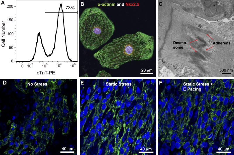
Methods: Cardiomyocytes derived from human-induced pluripotent stem cells were used to generate collagen-based bioengineered human cardiac tissue. Engineered tissue constructs were subjected to different mechanical stress and electric pacing conditions.

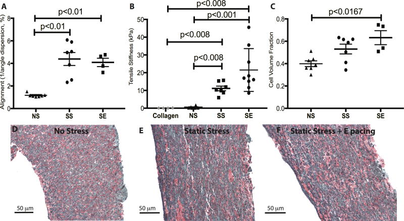
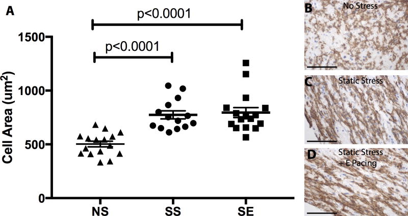
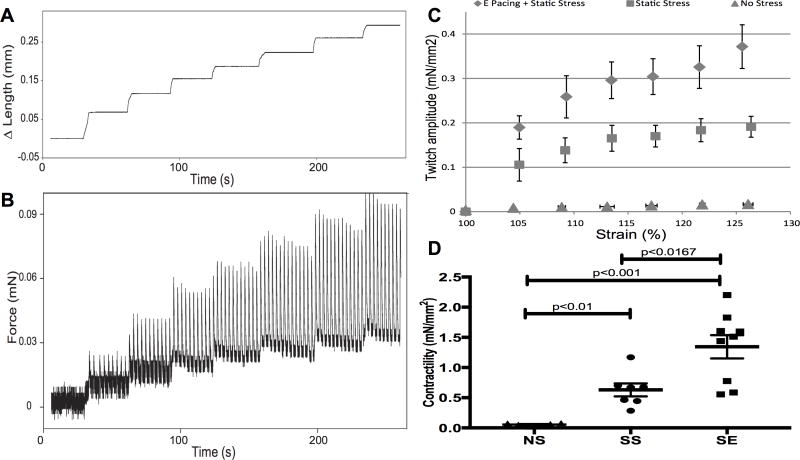
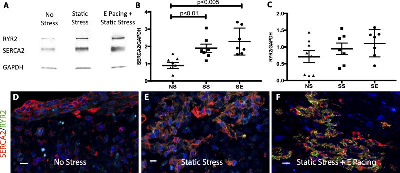
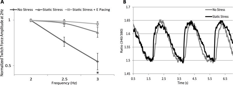
Results: The engineered human myocardium exhibits Frank-Starling-type force-length relationships. After 2 weeks of static stress conditioning, the engineered myocardium demonstrated increases in contractility (0.63±0.10 mN/mm2 vs 0.055±0.009 mN/mm2 for no stress), tensile stiffness, construct alignment, and cell size. Stress conditioning also increased SERCA2 (Sarco/Endoplasmic Reticulum Calcium ATPase 2) expression, which correlated with a less negative force-frequency relationship. When electric pacing was combined with static stress conditioning, the tissues showed an additional increase in force production (1.34±0.19 mN/mm2), with no change in construct alignment or cell size, suggesting maturation of excitation-contraction coupling. Supporting this notion, we found expression of RYR2 (Ryanodine Receptor 2) and SERCA2 further increased by combined static stress and electric stimulation.

Conclusions: These studies demonstrate that electric pacing and mechanical stimulation promote maturation of the structural, mechanical, and force generation properties of human-induced pluripotent stem cell-derived cardiac tissues.

Keywords: cardiomyocyte hypertrophy; electrical stimulation; human myocardium; stem cell; stress; tissue engineering.


Figures

Figure 1
Figure 1
Generation of high purity human iPS cell-derived constructs. A, Human cardiomyocytes were generated from the IMR90-iPS cell line at a purity of over 70% by cardiac troponin T (cTnT) flow cytometric analysis. B, The cardiomyocytes displayed robust sarcomeric organization by α-actinin immunostaining (green), as well as nuclear expression of the cardiomyocyte transcription factor Nkx2.5 (red). C, Primitive intercalated disc-like structures containing fascia adherens junctions and desmosomes (arrow) are present in the engineered heart tissue. Nuc: nucleus, Glyc: glycogen. Immunofluorescent staining for cardiac troponin T of iPS cells-derived human bioengineered cardiac tissues under NS, D, and SS, E, and SE, F, conditioning, shows greater alignment in the SS and SE tissue constructs.
Figure 2
Figure 2
Characterization of cell alignment and matrix remodeling. A, Constructs exposed to stress conditioning demonstrated significantly increased cell alignment compared to constructs without stress conditioning. Electrical pacing along with stress conditioning did not further promote the cell alignment. n=7 for NS, n=7 for SS, and n=4 for SE. B, The passive stiffness of constructs was measured by stretching constructs incrementally to 125% of slack length. Tensile stiffness was estimated from the slope of passive stress-strain relationship. The tensile stiffness of cell-free collagen matrix is 0.079 ± 0.041 kPa. Addition of cells increased the stiffness ~7-fold, stress conditioning by a further ~20-fold, and electrical pacing by an additional ~2-fold (NS: 0.47±0.22 kPa; SS: 11.13±1.17 kPa; SE: 21.51±4.02 kPa,). n=6 for NS, n=7 for SS, and n=9 for SE. C, Electrical pacing promotes ECM remodeling by increasing the cell volume fraction within the constructs (NS vs SS, p=0.0728, NS vs SE, p=0.0121, SS vs SE, P=0.3)). n=7 for NS, n=7 for SS, and n=4 for SE. D, E, and F, are representative Sirius Red/Fast Green stains of NS, SS and SE, respectively.
Figure 3
Figure 3
Stress conditioning increases cardiomyocyte hypertrophy. A, MYH7 positive area was measured to determine cardiomyocyte size from constructs in the different conditioning regimes. Cardiomyocyte size increased ~50% from NS vs. SS constructs, with no further increase in the ES group.(NS to SS, NS to SE, p<0.0001) B, C, and D, show MYH7 positive cells in constructs from NS, SS, and SE, respectively. n=16 for NS, n=14 for SS, and n=14 for SE. Hematoxylin counterstain denotes nuclei. The scale bar is 50 μm.
Figure 4
Figure 4
Stress conditioning and electrical stimulation increase contractility. Representative length (A) and force (B) traces demonstrate the response of a spontaneously contracting cardiac tissue construct to a series of stretches up to 125% of slack length. The amplitude of the isometric twitch force increases with increasing preparation length, in accordance with the Frank-Starling mechanism. C, Isometric twitch force amplitude measured at different preparation lengths is enhanced by 2 weeks of SS conditioning (triangles) in comparison to NS conditioning (triangles). Addition of electrical stimulation (diamonds) further increases contractility as show in (D). D, Contractility of constructs from the 3 stimulation conditions. Contractility is measured from the slope of the twitch force-strain curve, which is the active force development. The contractility of no stress constructs is 0.055±0.009 mN/mm2. Stress conditioning promotes the contractility 10-fold (0.63±0.10 mN/mm2) and addition of electrical pacing further enhances force development another 2-fold (1.34±0.19 mN/mm2). NS vs SS: p<0.01; SS vs SE: p<0.01. n=6 for NS, n=7 for SS, and n=9 for SE.
Figure 5
Figure 5
Increase in calcium handling protein expression by stress conditioning and electrical stimulation. A, Western blot of SR-related proteins, SERCA2 and RYR2, from constructs subjected to different conditioning regimes. B&C, Dot plot of quantified western blot data. Data are normalized to internal control (GAPDH). B, Compared to the NS control, SS conditioned constructs increase SERCA2 expression by 2-fold (NS vs SS, p<0.01) and SE-conditioned constructs by 2.5-fold (NS vs SE, p<0.005). C, An increasing RYR2 expression trend is observed from NS to SE constructs but did not reach statistical significance. n=8 for NS, n=7 for SS, and n=7 for SE. Representative immunostaining images of engineered cardiac tissues stained with SERCA2 (red) and RYR2 (green) from NS (D), SS (E) and SE (F) conditionings. The scale bar is 10 μm.
Figure 6
Figure 6
Force frequency relationship and calcium transient characteristics of engineered cardiac tissues. A, Engineered cardiac tissues were subjected to 2, 2.5, and 3 Hz pacing and the twitch amplitude at 2 Hz was used to normalize other pacing frequencies. Due to immature development of SR, the force frequency relations of these constructs are negative, but static stress and electrical pacing pre-conditioning were able to mitigate the effect (NS p=0.0047 for 2 Hz vs 3 Hz; SS p=0.09 for 2 Hz vs 3 Hz; SE p=0.8 for 2 Hz vs 3 Hz). n=5 for NS, n=11 for SS, and n=4 for SE. B, Representative calcium transient from NS (gray) and SS (black)-conditioned constructs under 0.5 Hz pacing.

Comment in

References

    1. Ogle BM, Bursac N, Domian I, Huang NF, Menasche P, Murry CE, Pruitt B, Radisic M, Wu JC, Wu SM, Zhang J, Zimmermann WH, Vunjak-Novakovic G. Distilling complexity to advance cardiac tissue engineering. Sci Transl Med. 2016;8:342ps13. - PMC - PubMed
    1. Jackman CP, Shadrin IY, Carlson AL, Bursac N. Human Cardiac Tissue Engineering: From Pluripotent Stem Cells to Heart Repair. Curr Opin Chem Eng. 2015;7:57–64. - PMC - PubMed
    1. Kurokawa YK, George SC. Tissue engineering the cardiac microenvironment: Multicellular microphysiological systems for drug screening. Adv Drug Deliv Rev. 2016;96:225–33. - PMC - PubMed
    1. Tzatzalos E, Abilez OJ, Shulda P, Wu JC. Engineered heart tissues and induced pluripotent stem cells: Macro- and microstructures for disease modeling, drug screening, and translational studies. Advanced Drug Delivery Reviews. 2016;96:234–244. - PMC - PubMed
    1. Feric NT, Radisic M. Strategies and Challenges to Myocardial Replacement Therapy. Stem Cells Translational Medicine. 2016;5:410–416. - PMC - PubMed