. 2016 Dec;15(12):3640-3652.
doi: 10.1074/mcp.M116.060491. Epub 2016 Oct 12.

Robust Label-free, Quantitative Profiling of Circulating Plasma Microparticle (MP) Associated Proteins

Affiliations

Robust Label-free, Quantitative Profiling of Circulating Plasma Microparticle (MP) Associated Proteins

Sophie Braga-Lagache et al. Mol Cell Proteomics. 2016 Dec.

Abstract

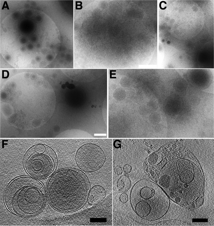
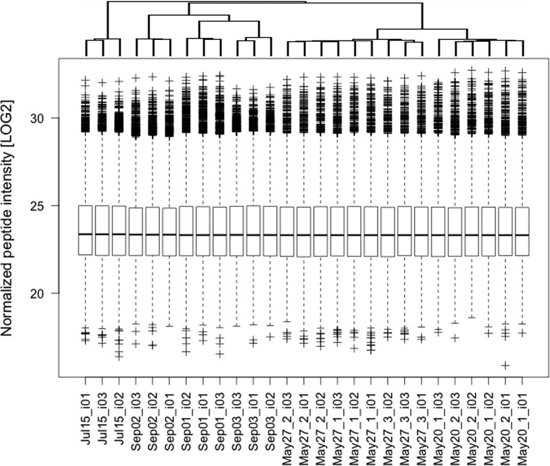
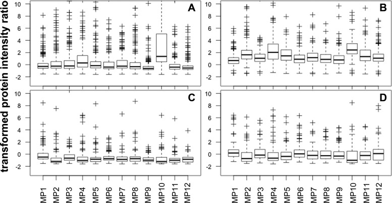
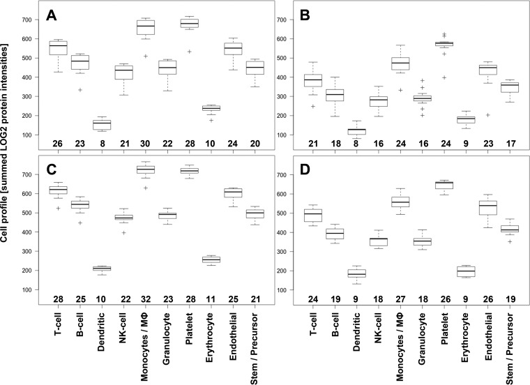
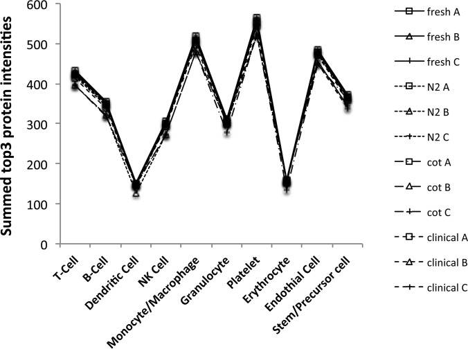
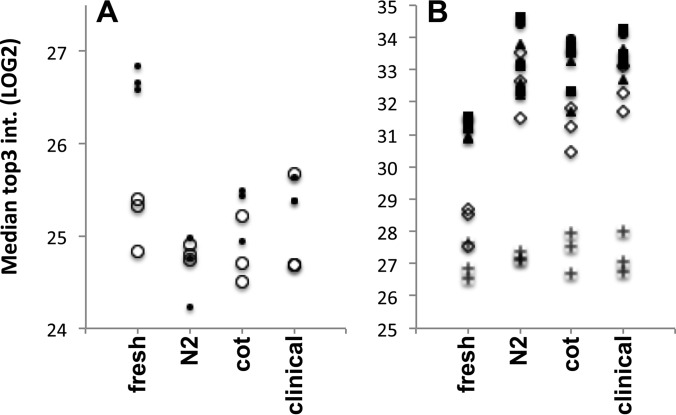
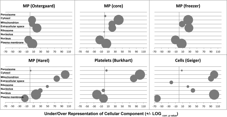
Cells of the vascular system release spherical vesicles, called microparticles, in the size range of 0.1-1 μm induced by a variety of stress factors resulting in variable concentrations between health and disease. Furthermore, microparticles have intercellular communication/signaling properties and interfere with inflammation and coagulation pathways. Today's most used analytical technology for microparticle characterization, flow cytometry, is lacking sensitivity and specificity, which might have led to the publication of contradicting results in the past.We propose the use of nano-liquid chromatography two-stage mass spectrometry as a nonbiased tool for quantitative MP proteome analysis.For this, we developed an improved microparticle isolation protocol and quantified the microparticle protein composition of twelve healthy volunteers with a label-free, data-dependent and independent proteomics approach on a quadrupole orbitrap instrument.Using aliquots of 250 μl platelet-free plasma from one individual donor, we achieved excellent reproducibility with an interassay coefficient of variation of 2.7 ± 1.7% (mean ± 1 standard deviation) on individual peptide intensities across 27 acquisitions performed over a period of 3.5 months. We show that the microparticle proteome between twelve healthy volunteers were remarkably similar, and that it is clearly distinguishable from whole cell and platelet lysates. We propose the use of the proteome profile shown in this work as a quality criterion for microparticle purity in proteomics studies. Furthermore, one freeze thaw cycle damaged the microparticle integrity, articulated by a loss of cytoplasm proteins, encompassing a specific set of proteins involved in regulating dynamic structures of the cytoskeleton, and thrombin activation leading to MP clotting. On the other hand, plasma membrane protein composition was unaffected. Finally, we show that multiplexed data-independent acquisition can be used for relative quantification of target proteins using Skyline software. Mass spectrometry data are available via ProteomeXchange (identifier PXD003935) and panoramaweb.org (https://panoramaweb.org/labkey/N1OHMk.url).


Conflict of interest statement

Disclosures: None.

Figures

Fig. 1.
Fig. 1.
Confirmation of MP by cryo-TEM. MP were isolated with three PBS washes without addition of SB and subjected to cryo-TEM. Panels A to E show five representative pictures at different magnification. Picture B depicts MP clumps. The white bars in B and D scale to 500 nm and the bright round shapes visible in each picture are holes in the carbon film with a diameter of 2 μm. Panels F and G show two slices through a volume of a tomographic composition from the same preparations showing multi-vesicular structures (F) and possibly encapsulated cellular organelles (G). The black bars scale to 200 μm.
Fig. 2.
Fig. 2.
Reproducibility in MP isolation and protein digestion. MP associated proteins were digested as described in the experimental procedures section and analyzed with a 60-min (May20 and May27 samples) or 90-min (July15 and Sep01 to Sep03 samples) ACN gradient, respectively. All the used 250 μl PFP aliquots were from the same donor, but prepared from two different blood samples being collected more than a year (May20, May27 and Jul15 samples) and 10 days (Sep01 to Sep03 samples) before use, respectively. The boxplots show the distributions of median normalized LOG2 peptide intensities as reported by MaxQuant software. The data is ordered according to hierarchical clustering (top). The first number after the date and an underscore relates to the sample number on the date of preparation. The _i0x (x = 1 to 3) relates to the LC-MS/MS run of this sample. None of the means were tested different from others (p = 0.05).
Fig. 3.
Fig. 3.
Quality check of label-free MP proteome quantification. Protein ratios were calculated between the 400 μl and 250 μl PFP input of 12 healthy donors (x axis) and transformed by subtraction of 1.6, the expected, theoretical ratio, in order to have a median ratio of zero (y axis). The upper panels A and B represent the ratios of cell membrane and cell membrane associated proteins, the lower panels C and D the ones of serum proteins. The panels on the left (A and C) show DDA MaxQuant top3 values, on the right (B and D) MaxQuant LFQ values, respectively. Only proteins identified in at least 12 out of 24 samples were considered for these figures.
Fig. 4.
Fig. 4.
Cell Profiling of MP origin. The log2 transformed cell marker protein intensities were summed (y axis) to create cell profiles of MP origin for all twelve plasma samples analyzed. The twelve values are represented as boxplots for the 250 μl PFP (top row, panels A and B) and 400 μl PFP input (bottom row, panels C and D) using the DDA top3 values on the left (panels A and C) and DIA-MSX3 Skyline data on the right (panels B and D). The numbers of protein matching to a cell type are given above the x axis for each boxplot.
Fig. 5.
Fig. 5.
Cell marker profiles of the twelve samples from the freezing study. A total of 31 cell marker proteins, representing the origin of MP (supplemental Table S5), were identified in the 12 MP isolates produced from plasma aliquots, which were frozen in different ways or processed without freezing (see experimental procedures section). The top3 protein intensities, represented as LOG2 values (y-axes), of cell specific markers (x axis) were summed. The capital letters A, B, C after the sample names depict the three technical replicates analyzed for each condition.
Fig. 6.
Fig. 6.
Differential recovery of proteins dependent on plasma freezing condition. An ANOVA test revealed 273 proteins that were significantly changed in abundance between the four tested plasma storage conditions (p ≤ 0.01 at FDR = 1%), with cytoskeleton proteins representing a major group. The median top3 intensities of the three replicates from cytoskeleton proteins were plotted in panel A, with small dots (●) representing proteins changed significantly and open circles (○) without a change in abundance, respectively. Panel B, the median top3 intensities of classical serum proteins with increased abundance after freezing (APOA2, APOH, CO3, CO8G, F13A, HRG, IGHM, KV110) are depicted as crosses. The three fibrin chains, alpha (●), beta (▴), and gamma (■), and fibronectin (♢) are shown individually.
Fig. 7.
Fig. 7.
Comparison of MP proteome composition with cell proteomes. The over- or under-representation of GO slim cellular component terms (x axis) were plotted for the published proteomes of MP by Østergaard et al. (18) and Harel et al. (according to supplemental Table S2D) (29), 11 different cell lines analyzed by Geiger et al. (30), platelets by Burkhart et al. (31), and two proteome sets from this study using the core proteome identified from twelve healthy donors and from one single donor used for the freezer study, respectively. The data sets are marked in the header of each bubble plot. GO terms differentiating best MP from cell proteomes are given on the y axis. The bubble size represents the relative contribution of proteins in each set and the bubbles are placed horizontally according to the Benjamini-Hochberg false discovery rate corrected p value in -LOG for over-representation and +LOG for under-representation.

References

    1. van der Pol E., Böing A. N., Gool E. L., and Nieuwland R. (2016) Recent developments in the nomenclature, presence, isolation, detection and clinical impact of extracellular vesicles. J. Thromb. Haemost 14, 48–56 - PubMed
    1. Boulanger C. M., Amabile N., and Tedgui A. (2006) Circulating microparticles: a potential prognostic marker for atherosclerotic vascular disease. Hypertension 48, 180–186 - PubMed
    1. Meziani F., Tesse A., and Andriantsitohaina R. (2008) Microparticles are vectors of paradoxical information in vascular cells including the endothelium: role in health and diseases. Pharmacol. Rep. 60, 75–84 - PubMed
    1. Chironi G. N., Boulanger C. M., Simon A., George F. D., Freyssinet J. M., and Tegui A. (2009) Endothelial microperticles in diseases. Cell Tissue Res. 335, 143–151 - PubMed
    1. Little K. M., Smalley M., Harthun N. L., and Ley K. (2010) The plasma microvesicle proteome. Semin. Thromb. Hemost 36, 845–856 - PubMed

LinkOut - more resources