. 2017 Mar;28(3):876-887.
doi: 10.1681/ASN.2016020132. Epub 2016 Oct 13.

Oxalobacter formigenes- Derived Bioactive Factors Stimulate Oxalate Transport by Intestinal Epithelial Cells

Affiliations

Oxalobacter formigenes- Derived Bioactive Factors Stimulate Oxalate Transport by Intestinal Epithelial Cells

Donna Arvans et al. J Am Soc Nephrol. 2017 Mar.

Abstract

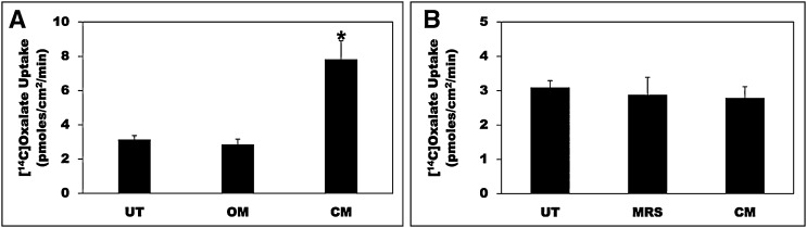
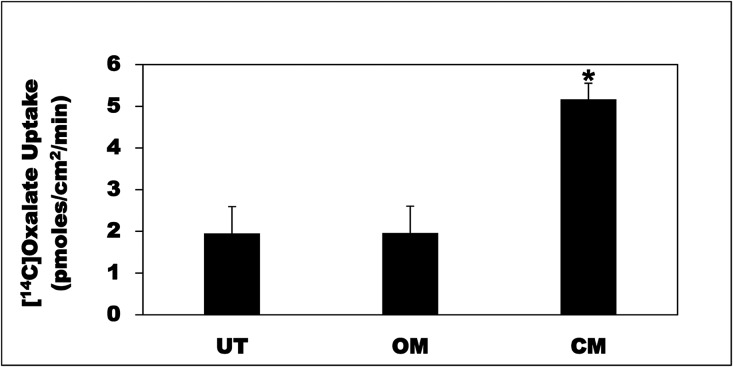
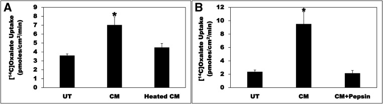
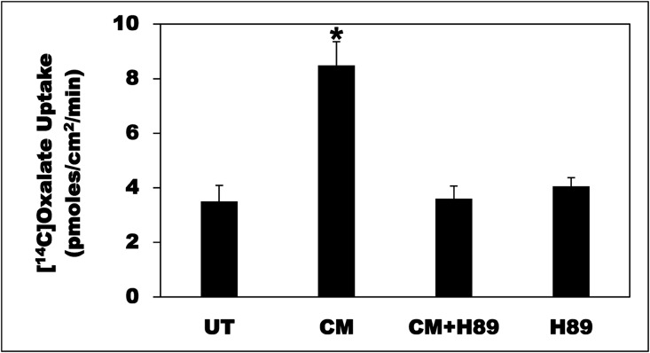
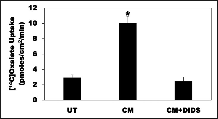
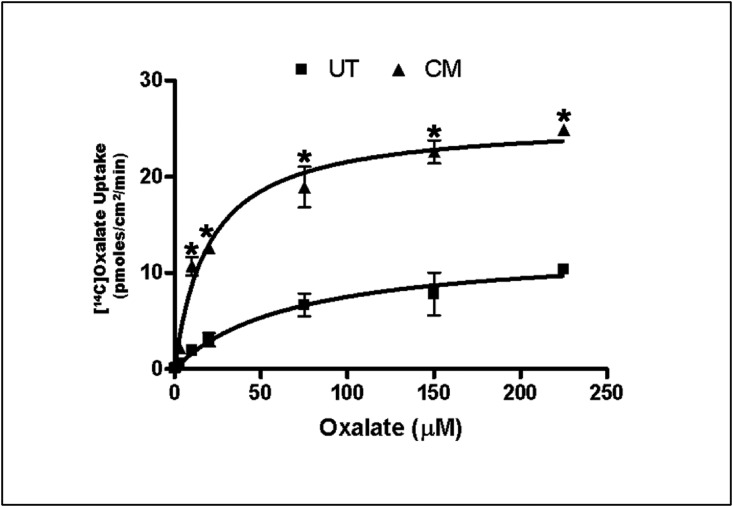
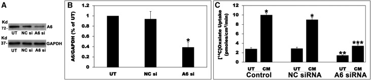
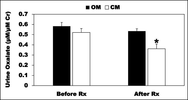
Hyperoxaluria is a major risk factor for kidney stones and has no specific therapy, although Oxalobacter formigenes colonization is associated with reduced stone risk. O. formigenes interacts with colonic epithelium and induces colonic oxalate secretion, thereby reducing urinary oxalate excretion, via an unknown secretagogue. The difficulties in sustaining O. formigenes colonization underscore the need to identify the derived factors inducing colonic oxalate secretion. We therefore evaluated the effects of O. formigenes culture conditioned medium (CM) on apical 14C-oxalate uptake by human intestinal Caco-2-BBE cells. Compared with control medium, O. formigenes CM significantly stimulated oxalate uptake (>2.4-fold), whereas CM from Lactobacillus acidophilus did not. Treating the O. formigenes CM with heat or pepsin completely abolished this bioactivity, and selective ultrafiltration of the CM revealed that the O. formigenes-derived factors have molecular masses of 10-30 kDa. Treatment with the protein kinase A inhibitor H89 or the anion exchange inhibitor 4,4'-diisothiocyano-2,2'-stilbenedisulfonic acid completely blocked the CM-induced oxalate transport. Knockdown of the oxalate transporter SLC26A6 also significantly restricted the induction of oxalate transport by CM. In a mouse model of primary hyperoxaluria type 1, rectal administration of O. formigenes CM significantly reduced (>32.5%) urinary oxalate excretion and stimulated (>42%) distal colonic oxalate secretion. We conclude that O. formigenes-derived bioactive factors stimulate oxalate transport in intestinal cells through mechanisms including PKA activation. The reduction in urinary oxalate excretion in hyperoxaluric mice treated with O. formigenes CM reflects the in vivo retention of biologic activity and the therapeutic potential of these factors.

Keywords: Oxalobacter formigenes; PKA; SLC26A6; intestinal oxalate transport; secreted bioactive factors.


Figures

Figure 1.
Figure 1.
O. formigenes CM stimulates oxalate uptake by C2 cells. (A) C2 cells were UT or were treated apically with OM or CM in the culture medium (1:50 dilution × 24 hours), and then 14C-oxalate uptake was measured as described in the Concise Methods. Values are means±SEM of 12 independent experiments each of which was done in triplicate. The CM significantly stimulated 14C-oxalate uptake (*P<0.001 for CM compared with UT and OM, by ANOVA). (B) C2 cells were UT or were treated apically with L. acidophilus growth medium (MRS) or CM in the culture medium (1:25 dilution × 24 hours), and then 14C-oxalate uptake was measured as described in the Concise Methods. Values are means±SEM of three independent experiments each of which was done in triplicate. L. acidophilus CM had no significant effect on 14C-oxalate uptake by C2 cells.
Figure 2.
Figure 2.
O. formigenes CM stimulates oxalate uptake by T84 cells. T84 cells were UT or were treated apically with OM or CM in the culture medium (1:50 dilution × 24 hours), and then 14C-oxalate uptake was measured as described in the Concise Methods. Values are means±SEM of three independent experiments each of which was done in triplicate. The CM significantly stimulated 14C-oxalate uptake (*P<0.05 and 0.01 for CM compared with UT and OM, respectively, by ANOVA).
Figure 3.
Figure 3.
Heat and pepsin treatment abolish the CM bioactivity. (A) C2 cells were UT or were treated apically with CM or a CM that was subjected to heat treatment (heated CM) in the culture medium (1:50 dilution × 24 hours), and then 14C-oxalate uptake was measured as described in the Concise Methods. Values are means±SEM of four independent experiments each of which was done in triplicate. The CM significantly stimulated 14C-oxalate uptake, an effect completely abolished by heat treatment (*P<0.01 and <0.05 for CM compared with UT and heated CM, respectively, by ANOVA). (B) C2 cells were UT or were treated apically with CM or a CM that was treated with pepsin (CM+Pepsin) in the culture medium (1:50 dilution × 24 hours), and then 14C-oxalate uptake was measured as described in the Concise Methods. Values are means±SEM of four independent experiments each of which was done in triplicate. The CM significantly stimulated 14C-oxalate uptake, an effect completely abolished by pepsin treatment (* P<0.01 for CM compared with UT and CM+Pepsin, by ANOVA).
Figure 4.
Figure 4.
Selective ultrafiltration shows the O. formigenes–derived factors to have molecular masses of 10–30 kDa. (A) Selective ultrafiltration of the CM using 10 kDa cutoff spin column. C2 cells were UT or were treated with CM, F, R, or the combined fractions (F+R) in the culture medium (1:50 dilution × 24 hours), and then 14C-oxalate uptake was measured as described in the Concise Methods. Values are means±SEM of five independent experiments each of which was done in duplicate or triplicate. The CM, the R, and the F+R significantly stimulated 14C-oxalate uptake (*P<0.001, <0.001, and <0.01 for CM compared with UT, F, and R; **P<0.01 for R compared with UT and F; and ***P<0.001, <0.001, and <0.01 for F+R compared with UT, F, and R, respectively, by ANOVA). (B) Selective ultrafiltration of the CM using 30 kDa cutoff spin column. C2 cells were UT or were treated with CM, F, R, or F+R in the culture medium (1:50 dilution × 24 hours), and then 14C-oxalate uptake was measured as described in the Concise Methods. Values are means±SEM of eight independent experiments each of which was done in triplicate. The CM, the F, and the F+R significantly stimulated 14C-oxalate uptake (*P<0.001 and <0.01 for CM compared with UT and R, respectively; ** P<0.05 for F compared with UT and R; and ***P<0.001 and <0.05 for F+R compared with UT and R, respectively, by ANOVA).
Figure 5.
Figure 5.
Lowering O. formigenes growth medium oxalate concentration leads to a CM with higher bioactivity. C2 cells were UT or treated with CM37.5, CM18.8, and CM9.4 (conditioned media prepared with growth medium oxalate concentration of 37.5 mM, 18.8 mM, and 9.4 mM, respectively) in the culture medium (1:50 dilution × 24 hours), and then 14C-oxalate uptake was measured as described in the Concise Methods. Values are means±SEM of three independent experiments each of which was done in triplicate. CM9.4 had significantly higher bioactivity compared with CM37.5 (*P<0.01 and <0.05 for CM9.4 compared with UT and CM37.5, respectively, by ANOVA).
Figure 6.
Figure 6.
The PKA inhibitor H89 completely blocked the CM-induced oxalate transport. C2 cells were UT or were treated apically with CM in the culture medium (1:50 dilution × 24 hours), and then 14C-oxalate uptake was measured as described in the Concise Methods. C2 cells were also treated apically with H89 (20 µM) for 30 minutes followed by the CM (1:50 dilution × 24 hours) with continued presence of H89 (CM+H89), or with H89 (20 µM) alone for 24.5 hours, and then 14C-oxalate uptake was similarly measured. Values are means±SEM of seven independent experiments each of which was done in triplicate. The CM significantly stimulated 14C-oxalate uptake, an effect completely blocked by H89 (*P<0.001 for CM compared with UT, CM+H89, and H89, by ANOVA).
Figure 7.
Figure 7.
The CM-induced oxalate transport is DIDS-sensitive. C2 cells were UT or were treated apically with CM in the culture medium (1:50 dilution × 24 hours), and then 14C-oxalate uptake was measured as described in the Concise Methods. CM-treated cells were treated apically with DIDS (100 µM) for 36 minutes (30 minutes immediately before the flux as well as during the 6 minute flux period) (CM+DIDS). Values are means±SEM of seven independent experiments each of which was done in triplicate. The CM significantly stimulated 14C-oxalate uptake, an effect completely abolished by DIDS (*P<0.001 for CM compared with UT and CM+DIDS, by ANOVA).
Figure 8.
Figure 8.
The CM has no effect on A1, A2, and A3 mRNA expression. C2 cells were UT or apically treated with CM in the culture medium (1:50 dilution × 24 hours), and then total RNA was isolated for real-time PCR analysis. Values are means±SEM of three independent experiments each of which was done in duplicate or triplicate. Relative A1, A2, and A6 mRNA expression levels were expressed as a percentage of UT normalized to GAPDH. The CM had no significant effect on A1, A2, and A6 mRNA expression levels.
Figure 9.
Figure 9.
The CM increases the Vmax and reduces the Km. C2 cells were UT or apically treated with CM in the culture medium (1:50 dilution × 24 hours), and then14C-oxalate uptake as a function of increasing 14C-oxalate concentration in the flux medium (0.3, 1, 3, 10, 20, 75, 150, and 225 µM) was assessed. Values are means±SEM of three independent experiments each of which was done in triplicate. The CM significantly stimulated 14C-oxalate uptake at each oxalate concentration (*P<0.03, 0.003, 0.003, 0.01, 0.002, 0.01, 0.01, and 0.001 for CM compared with UT at the above corresponding 14C-oxalate concentrations, by unpaired t test).
Figure 10.
Figure 10.
Silencing A6 greatly reduced the CM-induced stimulation of oxalate transport. (A) A representative Western blot analysis of total A6 protein expression. A6 protein expression was evaluated in C2 cell lysate (10 µg protein/lane: untransfected cells; NC si, C2 cells transfected with the NC si; A6 si, C2 cells transfected with the siRNA targeting A6). The lower half of the same blot was probed with an anti-GAPDH antibody to normalize loading of protein in each lane (lower panel). (B) Densitometry of immunoblot results. Western blot band density was quantified using ImageJ software. Values are means±SEM for six independent experiments of relative total A6 abundance to GAPDH and are presented as a percentage of the UT value. A6 siRNA knockdown significantly reduced A6 protein expression (*P<0.001 for A6 si compared with UT and NC si, by ANOVA). (C) Effect of A6 silencing on the CM-induced stimulation of 14C-oxalate uptake by C2 cells. Untransfected (Control), NC siRNA (transfected with the NC si), and A6 siRNA (transfected with the siRNA targeting A6) C2 cells were UT or were treated apically with the CM in the culture medium (1:50 dilution × 24 hours), and then 14C-oxalate uptake was measured as described in the Concise Methods. Values are means±SEM of eight independent experiments each of which was done in triplicate. Silencing A6 significantly reduced the CM-induced stimulation of oxalate transport by C2 cells (*P<0.001 for CM compared with UT in the Control and NC siRNA; **P<0.01 for UT in A6 siRNA compared with UT in Control and NC siRNA; ***P<0.001 for CM in A6 siRNA compared with CM in Control and NC siRNA, by ANOVA).
Figure 11.
Figure 11.
O. formigenes CM reduces urinary oxalate excretion in PH1 mice. The PH1 mice were given the CM or OM (100 µl twice daily as rectal enemas × 21 days; n=6–8) and urine was collected before (Before Rx) and after treatment (After Rx) for oxalate measurements. The CM significantly reduced urinary oxalate (adjusted for urinary creatinine [Cr]) excretion (*P<0.03 for CM compared with OM, by unpaired t test).

References

    1. Alexander RT, Hemmelgarn BR, Wiebe N, Bello A, Morgan C, Samuel S, Klarenbach SW, Curhan GC, Tonelli M; Alberta Kidney Disease Network : Kidney stones and kidney function loss: a cohort study. BMJ 345: e5287, 2012 - PMC - PubMed
    1. Coe FL, Evan A, Worcester E: Kidney stone disease. J Clin Invest 115: 2598–2608, 2005 - PMC - PubMed
    1. Curhan GC, Taylor EN: 24-h uric acid excretion and the risk of kidney stones. Kidney Int 73: 489–496, 2008 - PubMed
    1. Robertson WG, Peacock M: The cause of idiopathic calcium stone disease: hypercalciuria or hyperoxaluria? Nephron 26: 105–110, 1980 - PubMed
    1. Kleinman JG: Bariatric surgery, hyperoxaluria, and nephrolithiasis: a plea for close postoperative management of risk factors. Kidney Int 72: 8–10, 2007 - PubMed

LinkOut - more resources