. 2016:2016:1542786.
doi: 10.1155/2016/1542786. Epub 2016 Sep 25.

CYLD Promotes TNF- α-Induced Cell Necrosis Mediated by RIP-1 in Human Lung Cancer Cells

Affiliations

CYLD Promotes TNF- α-Induced Cell Necrosis Mediated by RIP-1 in Human Lung Cancer Cells

Xing Lin et al. Mediators Inflamm. 2016.

Abstract

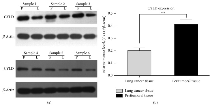
Lung cancer is one of the most common cancers in the world. Cylindromatosis (CYLD) is a deubiquitination enzyme and contributes to the degradation of ubiquitin chains on RIP1. The aim of the present study is to investigate the levels of CYLD in lung cancer patients and explore the molecular mechanism of CYLD in the lung cancer pathogenesis. The levels of CYLD were detected in human lung cancer tissues and the paired paracarcinoma tissues by real-time PCR and western blotting analysis. The proliferation of human lung cancer cells was determined by MTT assay. Cell apoptosis and necrosis were determined by FACS assay. The results demonstrated that low levels of CYLD were detected in clinical lung carcinoma specimens. Three pairs of siRNA were used to knock down the endogenous CYLD in lung cancer cells. Knockdown of CYLD promoted cell proliferation of lung cancer cells. Otherwise overexpression of CYLD induced TNF-α-induced cell death in A549 cells and H460 cells. Moreover, CYLD-overexpressed lung cancer cells were treated with 10 μM of z-VAD-fmk for 12 hours and the result revealed that TNF-α-induced cell necrosis was significantly enhanced. Additionally, TNF-α-induced cell necrosis in CYLD-overexpressed H460 cells was mediated by receptor-interacting protein 1 (RIP-1) kinase. Our findings suggested that CYLD was a potential target for the therapy of human lung cancers.


Conflict of interest statement

The authors have declared that no conflict of interests exists.

Figures

Figure 1
Figure 1
Lower levels of CYLD are detected in clinical lung carcinoma specimens. The surgical specimens including lung cancer tissues and the paired peritumoral tissues from the lung cancer patients were collected. (a) The expression levels of CYLD were detected by western blotting analysis. The data shown here was representative of six independent specimens. The housekeeping gene, β-actin, was used as an internal reference in the experiment. P, paracarcinoma tissue; L, lung adenocarcinoma tissue. (b) The levels of CYLD were detected by real-time PCR assay. Ratios of CYLD/β-actin were shown representing the relative mRNA levels in lung cancer patients. The data was shown as mean values ± SD. ∗∗ p < 0.01 compared with peritumoral tissue.
Figure 2
Figure 2
Knockdown of CYLD by siRNA promotes the growth of A549 and H460 cells. (a) The A549 cells were plated into 24-well plate. Eight hours later, three pairs of CYLD-siRNA were transfected into A549 cells. The transfected cells were cultured for 48 hours; the expression of CYLD was detected by western blotting analysis. (b) β-Actin was used as an internal reference and N.C. siRNA was used as the negative control. The expression level of CYLD was normalized to the expression of β-actin. ∗∗ p < 0.01, compared with the N.C. siRNA transfected group. (c) Cell viability was determined by MTT assay. The transfected A549 cells were cultured for 24 hours, 48 hours, and 72 hours, respectively, and the OD490 nm values were detected by MTT assay. ∗∗ p < 0.01, compared with N.C. siRNA group. (d) H460 cells were transfected with three pairs of siRNA specific to CYLD and cultured for 24 h, 48 h, and 72 h, respectively. The cell viability was determined by MTT assay. p < 0.05, ∗∗ p < 0.01, compared with N.C. siRNA group.
Figure 3
Figure 3
Transfection of pcDNA3.1(+)-CYLD plasmid increases CYLD expression in lung cancer cell lines. (a) The lung cancer cells A549 and H460 were transfected with the recombinated plasmid pcDNA3(+)/CYLD-flag and control plasmid pcDNA3.1(+) for 24 hours. The levels of CYLD were detected by in lung cancer cell lines western blotting analysis. (b) The images were captured by image J software and gray values were analyzed and shown in histogram. ∗∗ p < 0.01, compared with control plasmid. (c) A549 cells were transfected with CYLD-flag plasmid and cultured for 24 h, 48 h, and 72 h, respectively. The cell viability was determined by MTT assay. p < 0.05, ∗∗ p < 0.01, compared with the control plasmid transfected cells. (d) H460 cells were transfected with CYLD-flag plasmid for 24 h, 48 h, and 72 h. The cell viability was determined by MTT assay. p < 0.05, ∗∗ p < 0.01, compared with the control plasmid transfected cells.
Figure 4
Figure 4
Overexpression of CYLD-flag induces cell necrosis of H460 cells. (a) Induction of apoptosis and necrosis was determined by Annexin V-PI dural staining. The A549 stable cell lines with CYLD expression and the control cell line were plated into 12-well plate and treated with 10 ng/mL of TNF-α alone, 20 ng/mL of TNF-α combinated with 10 μM of a pancaspase inhibitor, z-VAD-fmk for 12 hours. The 2 × 10⁶ cells were collected and washed by PBS buffer. Annexin v-FITC/PI dual staining analysis was determined as the Material and Method. (b) The apoptosis rate of lung cells transfected with CYLD-flag or control plasmid was shown in histogram. p < 0.01, compared with control plasmid. ∗∗ p < 0.01, compared with N.C. siRNA group. (c) The necrosis rate was analyzed and shown in histogram. Values were mean ± SD. ∗∗ p < 0.01, compared with control plasmid.
Figure 5
Figure 5
Overexpression of CYLD enhances TNF-α-induced cell necrosis of lung cancer cells. (a) The necrosis was determined by FACS assay with PI staining. The CYLD-flag plasmid was used to transfect the lung cancer cell line H460 cells. The stable transfected cell line was screened as described in Material and Method. Increasing concentration of TNF-α, including 0 ng/mL, 1 ng/mL, 20 ng/mL, and 400 ng/mL, respectively, was used to treat the CYLD-flag plasmid transfected H460 cells. (b) The PI-positive rates of CYLD-flag transfected H460 cells were shown in histogram. ∗∗ p < 0.01, compared with pcDNA3.1(+) transfected H460 cells. (c) The CYLD-flag transfected cells were pretreated with a pancaspase inhibitor, z-VAD-fmk at the concentration of 10 μM for 6 hours, and different concentrations of TNF-α were used to treat H460 to induce cell necrosis. The PI-positive rates of H460 cells were shown in histogram. p < 0.05, ∗∗ p < 0.01, compared with the group without z-VAD-fmk.
Figure 6
Figure 6
TNF-α-induced cell necrosis in CYLD-overexpressed H460 cells is mediated by receptor-interacting protein 1 (RIP-1) kinase. (a) The stable cell line of CYLD-flag transfected H460 cells was plated into 6-well plate. The cells were cultured for 6 hours and transfected with two pairs of siRNA specific to RIP-1 for 48 hours. The levels of endogenous RIP-1 and CYLD were detected by western blotting analysis. β-Actin was used as internal reference gene in the present experiment. (b) The CYLD-overexpressed H460 cells were transfected with siRNA specific to RIP1 for 36 h and 48 h. The necrosis rate (PI-positive rate) was determined by FACS analysis. ∗∗ p < 0.01, compared with the cells transfected with N.C. siRNA. (c) The H460 cells were plated into 12-well plate and treated with 100 ng/mL of TNF-α in combination with or without 10 μM of z-VAD-fmk for 24 hours. Then, the cells were collected and PI-positive cell rate was determined as described in Material and Method. ∗∗ p < 0.01, compared with the group without z-VAD. (d) The CYLD-overexpressed H460 cells were plated into 6-well plate and treated with 100 ng/mL of TNF-α in combination with increasing concentrations of Nec-1 for 24 hours. The concentrations of Nec-1 were 0 μM, 0.1 μM, 1.0 μM, and 10 μM, respectively. The expression levels of RIP-1 were determined by western blotting analysis.

References

    1. Li X.-B., Gu J.-D., Zhou Q.-H. Review of aerobic glycolysis and its key enzymes—new targets for lung cancer therapy. Thoracic Cancer. 2015;6(1):17–24. doi: 10.1111/1759-7714.12148. - DOI - PMC - PubMed
    1. Ausborn N. L., Le Q. T., Bradley J. D., et al. Molecular profiling to optimize treatment in non-small cell lung cancer: a review of potential molecular targets for radiation therapy by the translational research program of the radiation therapy oncology group. International Journal of Radiation Oncology, Biology, Physics. 2012;83(4):e453–e464. doi: 10.1016/j.ijrobp.2012.01.056. - DOI - PubMed
    1. Keating G. M. Nivolumab: a review in advanced squamous non-small cell lung cancer. Drugs. 2015;75(16):1925–1934. doi: 10.1007/s40265-015-0492-9. - DOI - PubMed
    1. Cimas F. J., Callejas-Valera J., Pascual-Serra R., et al. MKP1 mediates chemosensitizer effects of E1a in response to cisplatin in non-small cell lung carcinoma cell. Oncotarget. 2015;6(42):44095–44107. doi: 10.18632/oncotarget.6574. - DOI - PMC - PubMed
    1. Eliyakin N., Postaci H., Baskin Y., Kozacioğlu Z. Small cell carcinoma of the urinary bladder: KIT and PDGFRA gene mutations. Rare Tumors. 2015;7(4, article 5982) doi: 10.4081/rt.2015.5982. - DOI - PMC - PubMed

MeSH terms

Substances