

A pediatric case of pituitary macroadenoma presenting with pituitary apoplexy and cranial nerve involvement: case report
- PMID: 27738402
- PMCID: PMC5047366
- DOI: 10.5152/TurkPediatriArs.2016.1945
A pediatric case of pituitary macroadenoma presenting with pituitary apoplexy and cranial nerve involvement: case report
Abstract
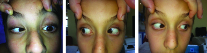
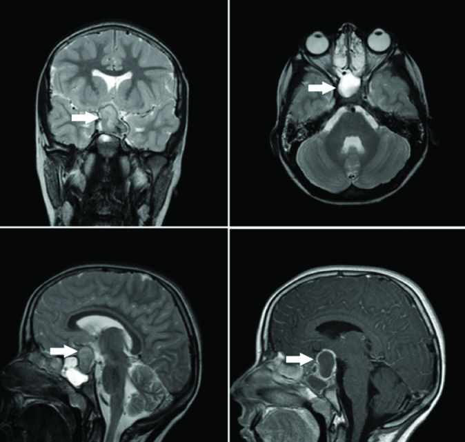
Pituitary adenomas usually arise from the anterior lobe of the pituitary gland and are manifested with hormonal disorders or mass effect. Mass effect usually occurs in nonfunctional tumors. Pituitary adenomas may be manifested with visual field defects or rarely in the form of total oculomotor palsy. Visual field defect is most frequently in the form of bitemporal hemianopsia and superior temporal defect. Sudden loss of vision, papilledema and ophthalmoplegia may be observed. Pituitary apoplexy is defined as an acute clinical syndrome characterized with headache, vomiting, loss of vision, ophthalmoplegia and clouding of consciousness. The problem leading to pituitary apoplexy may be decreased blood supply in the adenoma and hemorrhage following this decrease or hemorrhage alone. In this article, we present a patient who presented with fever, vomiting and sudden loss of vision and limited outward gaze in the left eye following trauma and who was found to have pituitary macroadenoma causing compression of the optic chiasma and optic nerve on the left side on cranial and pituitary magnetic resonance imaging.
Keywords: Adenoma; pituitary apoplexy; sudden loss of vision.
Figures
References
-
- Berkman ZM. Hipofiz adenomları. Türkiye Klinikleri J Surg Med Sci. 2007;3:152–64.
-
- Yılmaz M, İzmirli M, Yuca K, Mumcu Ç, Ünal Ö. Nasal ve nasofarengeal kitle olarak bulgu veren bir dev pituiter adenom olgusu. Tıp Araştırmaları Dergisi. 2006;4:45–7.
-
- Taylor M, Couto-Silva AC, Adan L, et al. Hypothalamic-pituitary lesions in pediatric patients: endocrine symptoms often precede neuro-ophthalmic presenting symptoms. J Pediatr. 2012;161:855–63. http://dx.doi.org/10.1016/j.jpeds.2012.05.014. - DOI - PubMed
-
- Erbaş T. Pituiter apopleksi. Turkiye Klinikleri J Surg Med Sci. 2006;2:28–30.
-
- Uğraş S. Hipofiz adenomlarında transsfenoidal mikrocerrahi. Uzmanlık Tezi, İstanbul. 2005. pp. 1–109.
Publication types
LinkOut - more resources
Full Text Sources
Other Literature Sources
