

Blue Laser Imaging-Bright Improves Endoscopic Recognition of Superficial Esophageal Squamous Cell Carcinoma
- PMID: 27738428
- PMCID: PMC5055998
- DOI: 10.1155/2016/6140854
Blue Laser Imaging-Bright Improves Endoscopic Recognition of Superficial Esophageal Squamous Cell Carcinoma
Abstract
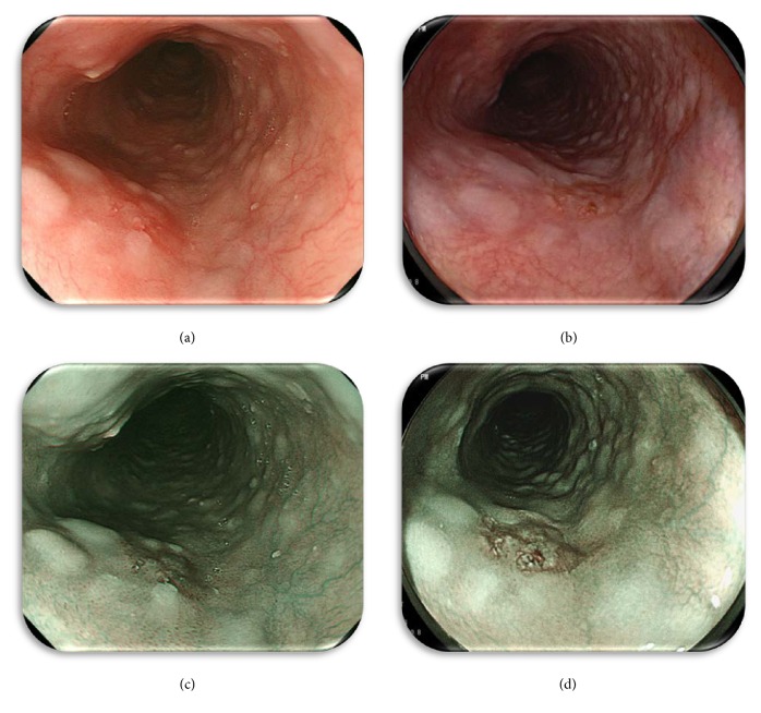
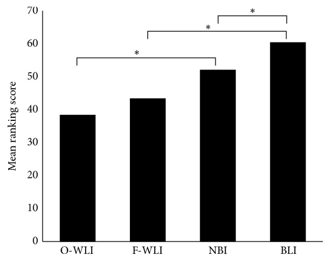
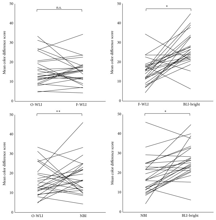
Background/Aims. The aim of this study was to evaluate the endoscopic recognition of esophageal squamous cell carcinoma (ESCC) using four different methods (Olympus white light imaging (O-WLI), Fujifilm white light imaging (F-WLI), narrow band imaging (NBI), and blue laser imaging- (BLI-) bright). Methods. We retrospectively analyzed 25 superficial ESCCs that had been examined using the four different methods. Subjective evaluation was provided by three endoscopists as a ranking score (RS) of each image based on the ease of detection of the cancerous area. For the objective evaluation we calculated the color difference scores (CDS) between the cancerous and noncancerous areas with each of the four methods. Results. There was no difference between the mean RS of O-WLI and F-WLI. The mean RS of NBI was significantly higher than that of O-WLI and that of BLI-bright was significantly higher than that of F-WLI. Moreover, the mean RS of BLI-bright was significantly higher than that of NBI. Furthermore, in the objective evaluation, the mean CDS of BLI-bright was significantly higher than that of O-WLI, F-WLI, and NBI. Conclusion. The recognition of superficial ESCC using BLI-bright was more efficacious than the other methods tested both subjectively and objectively.
Conflict of interest statement
Yoshito Itoh has an affiliation with a domination-funded department from Fujifilm Medical Co., Ltd. The other authors have no financial conflict of interests.
Figures

References
-
- Ozawa S., Tachimori Y., Baba H., et al. Comprehensive registry of esophageal cancer in Japan, 2002. Esophagus. 2010;7(1):7–22. doi: 10.1007/s10388-010-0228-6. - DOI
-
- Dawsey S. M., Fleischer D. E., Wang G.-Q., et al. Mucosal iodine staining improves endoscopic visualization of squamous dysplasia and squamous cell carcinoma of the esophagus in Linxian, China. Cancer. 1998;83(2):220–231. - PubMed
LinkOut - more resources
Full Text Sources
Other Literature Sources
