Clinical Trial
. 2017 Apr;31(4):872-881.
doi: 10.1038/leu.2016.282. Epub 2016 Oct 14.

Dynamic changes in the clonal structure of MDS and AML in response to epigenetic therapy

Affiliations
Clinical Trial

Dynamic changes in the clonal structure of MDS and AML in response to epigenetic therapy

G L Uy et al. Leukemia. 2017 Apr.

Abstract

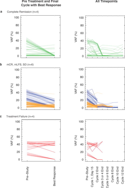
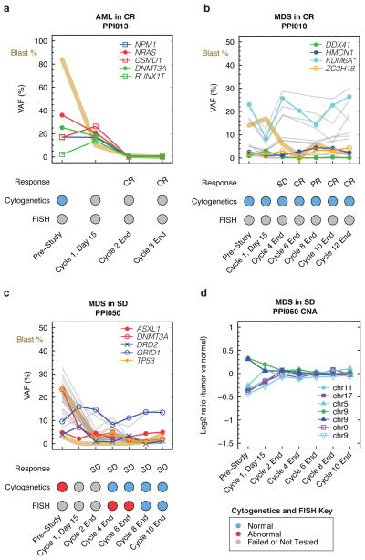
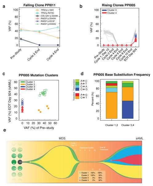
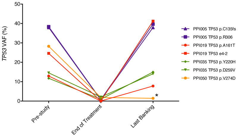
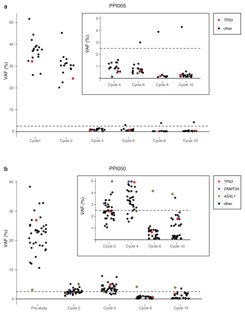
Traditional response criteria in myelodysplastic syndrome (MDS) and acute myeloid leukemia (AML) are based on bone marrow morphology and may not accurately reflect clonal tumor burden in patients treated with non-cytotoxic chemotherapy. We used next-generation sequencing of serial bone marrow samples to monitor MDS and AML tumor burden during treatment with epigenetic therapy (decitabine and panobinostat). Serial bone marrow samples (and skin as a source of normal DNA) from 25 MDS and AML patients were sequenced (exome or 285 gene panel). We observed that responders, including those in complete remission (CR), can have persistent measurable tumor burden (that is, mutations) for at least 1 year without disease progression. Using an ultrasensitive sequencing approach, we detected extremely rare mutations (equivalent to 1 heterozygous mutant cell in 2000 non-mutant cells) months to years before their expansion at disease relapse. While patients can live with persistent clonal hematopoiesis in a CR or stable disease, ultimately we find evidence that expansion of a rare subclone occurs at relapse or progression. Here we demonstrate that sequencing of serial samples provides an alternative measure of tumor burden in MDS or AML patients and augments traditional response criteria that rely on bone marrow blast percentage.

Trial registration: ClinicalTrials.gov NCT00691938.


Conflict of interest statement

Conflict of interest statement: GLU has received compensation as a consultant for Novartis. The remaining authors report no relevant conflicts of interest.

Figures

Figure 1
Figure 1. Heatmap of molecular and clinical findings
(a) Distribution of mutations in 25 patients (10 MDS, 15 AML) with at least one mutation in 16 genes or pathways in samples from any time-point. Each column represents an individual patient sample and each row represents a gene with a mutation. Mutations are indicated by colored cells and gene groups/families are indicated at the left. (b) The number of mutations in each gene present in pre-study samples is listed. Splice mutations include the splice acceptor and donor dinucleotides. (c) Number of coding mutations detected in the pre-study samples (excluding silent mutations). RMG, recurrently mutated genes in AML and MDS; Complex cytogenetics, ≥ 3 clonal abnormalities; Complete Remission, CR/CRc/CRi; Non-evaluable, received < 2 cycle of treatment; TF, transcription factors; PPI, positive patient identifier
Figure 2
Figure 2. Dynamic changes in mutation VAFs during treatment
Mutation variant allele fraction (VAF) in paired pre-study and time of best response samples from patients that achieved a complete remission (a) versus those that did not (b and c) (left panels). Each line represents a mutation that occurred in a diploid part of the genome. Serial mutation VAFs are displayed in the right panel. Stable disease (SD, n=2, blue), marrow leukemia free state (mLFS, n=2, orange), or marrow complete remission (mCR, n=2, orange). VAF, variant allele fraction.
Figure 3
Figure 3. Persistence of mutation VAFs in complete remission and stable disease
(a) An AML patient with normal cytogenetics (PPI013) achieved a CR at the end of cycle 2. The blast % decreased at cycle 1 day 15 prior to a decrease in 5 mutation VAFs, and normalized by the end of cycle 2. The molecular and morphologic response was similar in this patient. (b) An MDS patient with normal cytogenetics (PPI010) achieved a CR at the end of cycle 6 and 12. Mutations VAFs remained detectable throughout treatment, including a mutation in KDM6A that was present with a copy number adjusted VAF ~20–30% (i.e., ~40–60% of cells harboring a mutation). The molecular and morphologic response was discordant in this patient with a normal karyotype. (c) An MDS patient with abnormal cytogenetics (PPI050) had persistent stable disease throughout treatment. Cytogenetics and FISH normalized by cycle 8. While there was an initial decrease in mutations VAFs, they remained detectable throughout treatment. A mutation in ASXL1 was unchanged during treatment and may represent a clone that is independent of the malignant clone (i.e., clonal hematopoiesis of indeterminate significance). The molecular and morphologic response was similar and the patient remained with cytopenias despite have a low level of detectable mutations. (d) Copy number alterations detected by RMG sequencing for PPI050 are shown. Copy number gains have a log2 ratio of tumor versus normal >0 and losses <0. The altered regions are not detected by the end of cycle 2, including del(5). CR, complete remission; SD, stable disease; PR, partial remission; CNA, copy number alterations; VAF, variant allele fraction; FISH, fluorescent in situ hybridization; *copy number adjusted VAF.
Figure 4
Figure 4. Differential sensitivity of tumor clones during treatment
(a) An AML patient (PPI011) with treatment failure harbors a subclone defined by three somatic RAD21 mutations (colored lines) that become undetectable by the end of treatment (i.e., falling clone). Additional mutations (grey) define the founding clone that is treatment-resistant and contains two TP53 mutations. (b) An MDS patient who achieved CR at cycle 4 progressed to secondary AML (sAML) 924 days after the end of treatment on this study. Two subclones (red and blue) emerge and are detectable in the sAML sample. These subclones were not detected at pre-study or during the treatment cycles (i.e., rising clones). (c) The mutation VAFs from MDS patient PPI005 are shown at the pre-study MDS stage and after progression to sAML at 924 days after the end of treatment. Mutation VAFs were adjusted for chromosomal copy number. Unsupervised clustering of individual mutations identified four distinct mutation clusters representing clones, two of which (red and blue) are specific to the sAML sample. (d) Spectrum of single base substitutions in cluster 1 and 2 (present in initial pre-study MDS) versus clusters 3 and 4 (detectable in sAML). The sAML-specific clusters show a greater proportion of C-G transversions that are associated with decitabine treatment, suggesting that some mutations are caused by the treatment. (e) For patient PPI005, the tumor phylogeny was inferred using the clonevol package (https://github.com/hdng/clonevol, manuscript in preparation). Two models are possible, differing only in whether subclone 4 is derived from subclone 3 or 4 (Supplementary Figure 6). The model assigned higher likelihood was used to produce the above plot summarizing the clonal evolution from the MDS stage to the sAML. Residual non-mutant normal cells are not depicted at MDS or sAML time-points (i.e., percentage of cells with cluster mutations represents tumor cells only). Cells in clone 1 contain cluster 1 mutations. Clone 1 (green) is the founding clone and is present in nearly all bone marrow cells at MDS and sAML time points; clone 2 (yellow) is similarly present in almost all cells at the MDS pre-study time-point, but is present in only 33% of cells in the secondary AML time-point. Clones 3 and 4 (blue and red) are not detected in the pre-study MDS sample but emerge at sAML and are present in 60% and 32% of bone marrow cells, respectively. EOT, end of treatment. VAF, variant allele fraction.
Figure 5
Figure 5. TP53 mutation VAFs decrease during treatment
Four patients harbored somatic mutations inTP53 with VAFs >5% in the pre-study or cycle 1 time-point, completed at least 2 cycles of treatment, and had no evidence of TP53 loss of heterozygosity. These patients showed a mean VAF decrease of 26.5-fold at the end of treatment. All 4 patients had follow-up bone marrows obtained after completing the study, and 3 of 4 showed a subsequent increase in TP53 VAFs; the remaining patient (PPI050) continued on single agent decitabine after completing the study (indicated by *). Mutations are indicated in standard p-syntax.
Figure 6
Figure 6. Persistence of rare tumor cells are detected using ultra-deep error corrected sequencing
(a) An MDS patient (PPI005) achieved CR at the end of cycle 4, consistent with absence of mutation detection by standard sequencing platforms (conservative sensitivity indicated by the dashed line at 2% VAF). Somatic mutations were detected at all treatment time-points using ultra-deep error corrected sequencing and high sequence coverage, including during CR when many mutations are present with VAFs <1% (inset panel). Canonical gene mutations are indicated by colored circles. (b) An MDS patient with stable disease (no CR) shows similar persistence of detectable somatic mutations using ultra-deep error corrected sequencing across all treatment time-points, including those with VAFs <1% (inset). Mutations were detectable at cycles 8 and 10 when cytogenetics and FISH were negative. The ASXL1 mutation (green) does not change with treatment and is likely present in a nonmalignant clone distinct from the founding clone. VAF, variant allele fraction.

References

    1. Cashen AF, Schiller GJ, O’Donnell MR, DiPersio JF. Multicenter, phase II study of decitabine for the first-line treatment of older patients with acute myeloid leukemia. J Clin Oncol. 2009 Feb 1;28(4):556–561. - PubMed
    1. Silverman LR, Demakos EP, Peterson BL, Kornblith AB, Holland JC, Odchimar-Reissig R, et al. Randomized controlled trial of azacitidine in patients with the myelodysplastic syndrome: a study of the cancer and leukemia group B. J Clin Oncol. 2002 May 15;20(10):2429–2440. - PubMed
    1. Kantarjian H, Issa JP, Rosenfeld CS, Bennett JM, Albitar M, DiPersio J, et al. Decitabine improves patient outcomes in myelodysplastic syndromes: results of a phase III randomized study. Cancer. 2006 Apr 15;106(8):1794–1803. - PubMed
    1. Dombret H, Seymour JF, Butrym A, Wierzbowska A, Selleslag D, Jang JH, et al. International phase 3 study of azacitidine vs conventional care regimens in older patients with newly diagnosed AML with >30% blasts. Blood. 2015 Jul 16;126(3):291–299. - PMC - PubMed
    1. Fenaux P, Mufti GJ, Hellstrom-Lindberg E, Santini V, Gattermann N, Germing U, et al. Azacitidine prolongs overall survival compared with conventional care regimens in elderly patients with low bone marrow blast count acute myeloid leukemia. J Clin Oncol. 2010 Feb 1;28(4):562–569. - PubMed

Publication types

MeSH terms

Substances

Associated data