. 2016 Nov 15;7(46):74473-74483.
doi: 10.18632/oncotarget.12578.

7-Ketocholesterol induces ATM/ATR, Chk1/Chk2, PI3K/Akt signalings, cytotoxicity and IL-8 production in endothelial cells

Affiliations

7-Ketocholesterol induces ATM/ATR, Chk1/Chk2, PI3K/Akt signalings, cytotoxicity and IL-8 production in endothelial cells

Mei-Chi Chang et al. Oncotarget. .

Abstract

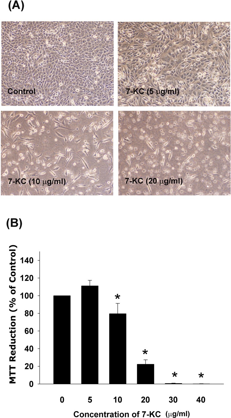
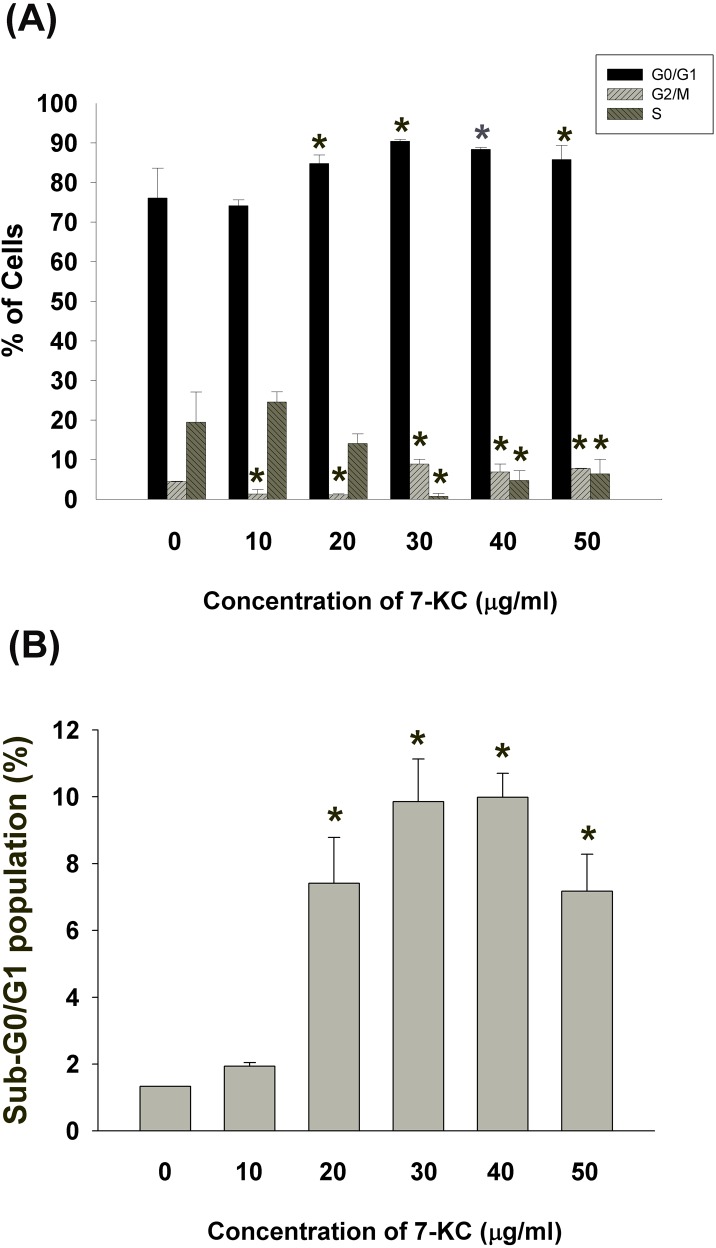
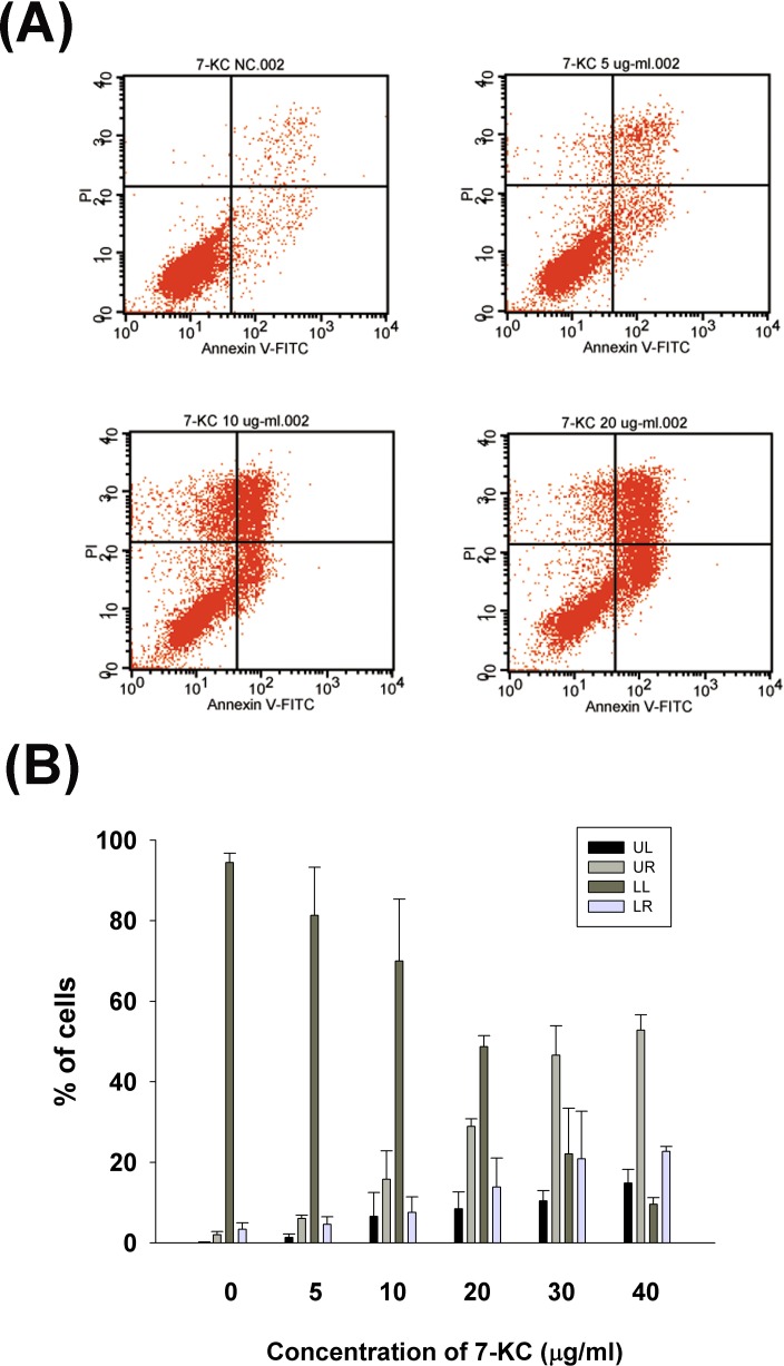
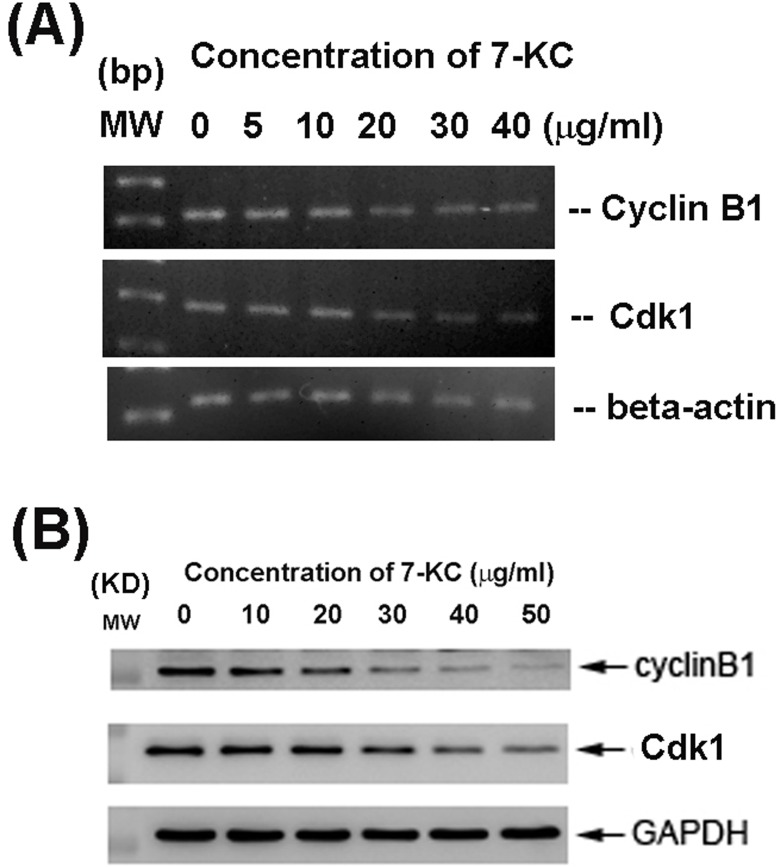
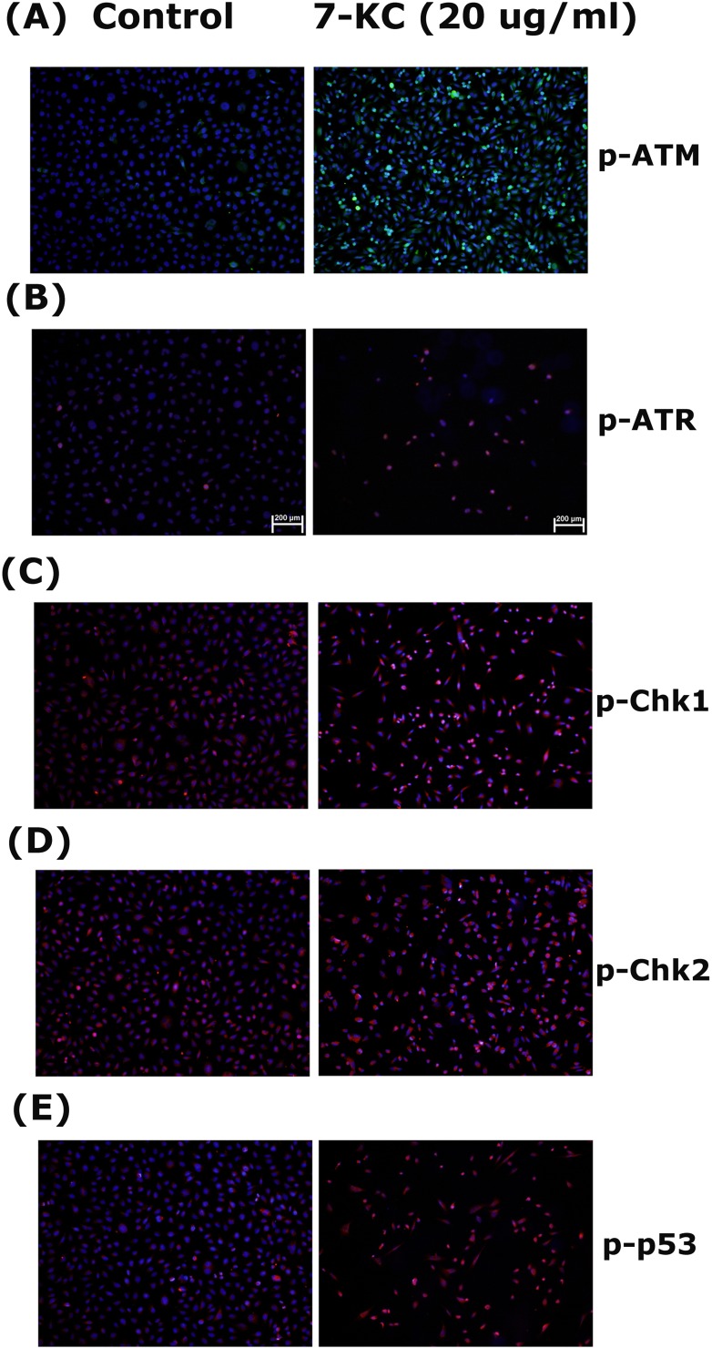
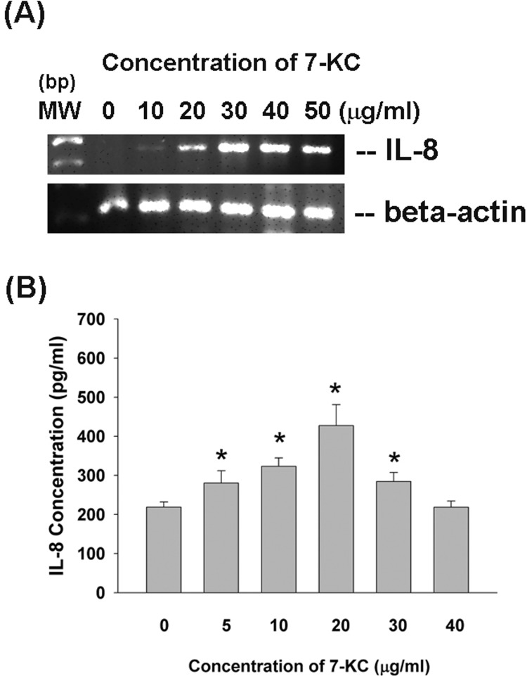
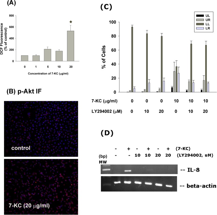
Cardiovascular diseases (atherosclerosis, stroke, myocardiac infarction etc.) are the major systemic diseases of elder peoples in the world. This is possibly due to increased levels of oxidized low-density lipoproteins (oxLDLs) such as 7-ketocholesterol (7-KC) and lysophosphatidylcholine (LPC) that damage vascular endothelial cells, induce inflammatory responses, to elevate the risk of cardiovascular diseases, Alzheimer's disease, and age-related macular degeneration. However the toxic effects of 7-KC on endothelial cells are not known. In this study, 7-KC showed cytotoxicity to endothelial cells at concentrations higher than 10 µg/ml. 7-KC stimulated ATM/Chk2, ATR-Chk1 and p53 signaling pathways in endothelial cells. 7-KC also induced G0/G1 cell cycle arrest and apoptosis with an inhibition of Cyclin dependent kinase 1 (Cdk1) and cyclin B1 expression. Secretion and expression of IL-8 in endothelial cells were stimulated by 7-KC. 7-KC further induced intracellular ROS production as shown by increase in DCF fluorescence and Akt phosphorylation. LY294002 attenuated the 7-KC-induced apoptosis and IL-8 mRNA expression of endothelial cells. These results indicate that oxLDLs such as 7-KC may contribute to the pathogenesis of atherosclerosis, thrombosis and cardiovascular diseases by induction of endothelial damage, apoptosis and inflammatory responses. These events are associated with ROS production, activation of ATM/Chk2, ATR/Chk1, p53 and PI3K/Akt signaling pathways.

Keywords: Gerotarget; apoptosis; atherosclerosis; cytotoxicity; endothelial cells; inflammation.


Conflict of interest statement

CONFLICTS OF INTERESTS

The authors declare no conflict of interest for this submission.

Figures

Figure 1
Figure 1. Cytotoxicity of 7-KC (10-40 μg/ml) to endothelial cells after 3 days of exposure
A. Morphologic changes of EAHY endothelial cells after exposure to different concentrations of 7-KC, B. Cyotoxicity of 7-KC to endothelial cells as analyzed by MTT. Results were expressed as Mean ± SE (n = 6). *denotes statistically significant difference (p < 0.05) when compared with solvent control.
Figure 2
Figure 2. Effect of 7-KC (10-50 μg/ml) on cell cycle progression and apoptosis of endothelial cells
A. Effect of 7-KC on cell cycle distribution of endothelial cells as analyzed by Modifit Software, B. Effect of 7-KC on sub-G0/G1 population of endothelial cells was analyzed by Cell Quest program. Results were expressed as Mean ± SE (n = 3).
Figure 3
Figure 3. Effect of 7-KC (5-40 μg/ml) on apoptosis of endothelial cells as analyzed by PI and annexin V dual fluorescent flow cytometry
A. One representative flow cytometry picture was shown. LL (lower left): viable cells, UL (upper left): necrotic cells, LR (lower right): pro-apoptotic cells, UR (upper right): apoptotic cells, B. Quantitative analysis of PI + annexin V flow cytometric analysis. Results were expressed as Mean ± SE (n = 3).
Figure 4
Figure 4. Effect of 24-h exposure to 7-KC on cell cycle-related Cdk1 and cyclin B1 mRNA and protein expression of endothelial cells
A. mRNA expression of Cdk1 and cyclin B1 as analyzed by PCR. Beta-actin expression was used as control. MW (molecular weight - base pairs [bp]) B. Cdk1 and cyclin B1 protein expression as analyzed by western blotting. MW (molecular weight, KD), Expression of beta-actin and GAPDH was used as control for PCR and western blot, respectively. One representative RT-PCR and western blotting result was shown.
Figure 5
Figure 5. Stimulation of p-ATM, p-ATR, p-Chk1, p-Chk2 and p-p53 expression by 7-KC (20 μg/ml) to endothelial cells
EAHY endothelial cells were exposed to solvent control and 20 μg/ml of 7-KC for 24 hours. Immunofluorescent (IF) microscopic observation was done to evaluate the expression of A. p-ATM, B. p-ATR, C. p-Chk1, D. p-Chk2 and E. p-p53 in endothelial cells. One representative IF picture was shown. (blue - DAPI, red or green - target proteins, p-ATM, p-ATR, p-Chk1, p-Chk2, p-p53)
Figure 6
Figure 6. Effect of 7-KC on IL-8 expression and production
EAHY endothelial cells were exposed to 7-KC. A. Total RNA was isolated and subjected to RT-PCR analysis of IL-8 expression, B. Culture medium was collected and used for measurement of IL-8 secretion in endothelial cells. Results were expressed as Mean ± SE (n = 5).
Figure 7
Figure 7. Effect of 7-KC on cellular ROS levels and Akt activation
A. 7-KC (10-40 μg/ml) induced ROS production as shown by increase in cellular DCF fluorescence. Results were expressed as Mean ± SE (% of control, n = 3), B. 7-KC stimulate Akt phosphorylation as shown by increase in cellular p-Akt fluorescence (blue - DAPI, red - p-Akt). One representative immunofluorescent study was shown. C. LY294002 attenuated the 7-KC-induced apoptosis of endothelial cells as analyzed by PI+Annexin V dual staining flow cytometry. Results were expressed as percentage (% of control, n = 6) of cell residing in UL, LL, UR and LR area of flow cytometry chart. D. LY294002 prevented the 7-KC-induced IL-8 mRNA expression of endothelial cells.

References

    1. Bourdon E, Loreau N, Davignon J, Bernier L, Blache D. Involvement of oxysterols and lysophosphatidylcholine in the oxidized LDL-induced impairment of serum albumin synthesis by HEPG2 cells. Arterioscler Thromb Vasc Biol. 2000;20:2643–2650. - PubMed
    1. Hughes H, Mathews B, Lenz ML, Guyton JR. Cytotoxicity of oxidized LDL to porcine aortic smooth muscle cells is associated with the oxysterols 7-ketocholesterol and 7-hydroxycholesterol. Arterioscler Thromb. 1994;14:1177–1185. - PubMed
    1. Lyon MA, Brown AJ. 7-Ketocholesterol. Int J Biochem Cell Biol. 1999;31:369–375. - PubMed
    1. Rodriguez IR, Larrayoz IM. Cholesterol oxidation in the retina: implications of 7KCh formation in chronic inflammation and age-related macular degeneration. J Lipid Res. 2010;51:2847–2862. - PMC - PubMed
    1. Gramajo AL, Zacharias LC, Neekhra A, Luthra S, Atilano SR, Chwa M, Brown DJ, Kuppermann BD, Kenney MC. Mitochondrial DNA damage induced by 7-ketocholesterol in human retina pigment epithelial cells in vitro. Invest Ophthalmol Vis Sci. 2010;51:1164–1170. - PubMed

MeSH terms