Review
. 2016 Sep 29;3(1):53-75.
doi: 10.1146/annurev-virology-100114-055104.

The Strange, Expanding World of Animal Hepaciviruses

Affiliations
Review

The Strange, Expanding World of Animal Hepaciviruses

Alex S Hartlage et al. Annu Rev Virol. .

Abstract

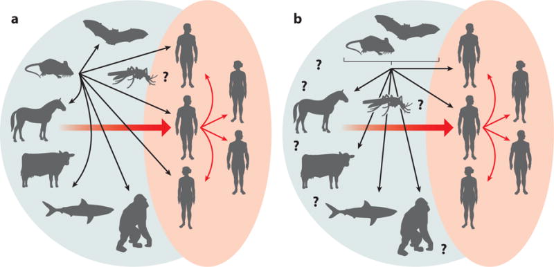
Hepaciviruses and pegiviruses constitute two closely related sister genera of the family Flaviviridae. In the past five years, the known phylogenetic diversity of the hepacivirus genera has absolutely exploded. What was once an isolated infection in humans (and possibly other primates) has now expanded to include horses, rodents, bats, colobus monkeys, cows, and, most recently, catsharks, shedding new light on the genetic diversity and host range of hepaciviruses. Interestingly, despite the identification of these many animal and primate hepaciviruses, the equine hepaciviruses remain the closest genetic relatives of the human hepaciviruses, providing an intriguing clue to the zoonotic source of hepatitis C virus. This review summarizes the significance of these studies and discusses current thinking about the origin and evolution of the animal hepaciviruses as well as their potential usage as surrogate models for the study of hepatitis C virus.

Keywords: animal models; hepatitis C virus; hepegivirus; pegivirus; virome; virus evolution.


Figures

Figure 1
Figure 1
Timeline of discoveries related to the animal hepaciviruses. Abbreviations: BHV, bat hepacivirus; BovHepV, bovine hepacivirus; CHV, canine hepacivirus; GBV-B, GB virus B; GHV, guereza hepacivirus; HCV, hepatitis C virus; NPHV, nonprimate hepacivirus; RHV, rodent hepacivirus;WLSV, Wenling shark virus.
Figure 2
Figure 2
Phylogenetic analysis of helicase gene (motifs I to IV) of representative members of (a) all four genera of the family Flaviviridae plus newly identified viruses and (b) the two genera Hepacivirus and Pegivirus. The evolutionary history was inferred using the maximum likelihood method based on the Le and Gascuel 2008 model (112). The tree with the highest log likelihood (−47,019.0830) is shown. The percentage of trees in which the associated taxa clustered together is shown next to each branch. Initial trees for the heuristic search were obtained automatically by applying Neighbor-Join (NJ) and BIONJ algorithms to a matrix of pairwise distances estimated by using a JTT model and then selecting the topology with superior log likelihood value. A discrete gamma distribution was used to model evolutionary rate differences among sites (five categories, +G, parameter = 1.7148). The rate variation model allowed for some sites to be evolutionarily invariable (+I, 3.9855% sites). The tree is drawn to scale, with branch lengths measured in number of substitutions per site. The analysis involved 96 amino acid sequences. There were a total of 276 positions in the final data set. Evolutionary analyses were conducted in MEGA7 (113).
Figure 2
Figure 2
Phylogenetic analysis of helicase gene (motifs I to IV) of representative members of (a) all four genera of the family Flaviviridae plus newly identified viruses and (b) the two genera Hepacivirus and Pegivirus. The evolutionary history was inferred using the maximum likelihood method based on the Le and Gascuel 2008 model (112). The tree with the highest log likelihood (−47,019.0830) is shown. The percentage of trees in which the associated taxa clustered together is shown next to each branch. Initial trees for the heuristic search were obtained automatically by applying Neighbor-Join (NJ) and BIONJ algorithms to a matrix of pairwise distances estimated by using a JTT model and then selecting the topology with superior log likelihood value. A discrete gamma distribution was used to model evolutionary rate differences among sites (five categories, +G, parameter = 1.7148). The rate variation model allowed for some sites to be evolutionarily invariable (+I, 3.9855% sites). The tree is drawn to scale, with branch lengths measured in number of substitutions per site. The analysis involved 96 amino acid sequences. There were a total of 276 positions in the final data set. Evolutionary analyses were conducted in MEGA7 (113).
Figure 3
Figure 3
Genome organization and polyprotein cleavage map of hepaciviruses, pegiviruses, and hepegiviruses. The complete genome encodes a polyprotein that is co- and posttranslationally cleaved into individual viral proteins. The structural proteins include the core protein (C) and envelope glycoproteins (E1 and E2), and the nonstructural proteins are NS2, NS3, NS4A, NS4B, NS5A, and NS5B. Classical pegiviruses (human and simian pegiviruses) do not have a recognizable C protein. Structural proteins are cleaved by cellular signal peptidases (black triangles), and NS2-NS3 cleavage is accomplished by the NS2-NS3 autoprotease (empty triangle). The remaining nonstructural proteins are cleaved by the NS3-NS4A protease complex (gray triangles). Hepegiviruses encode a protein termed Y that is similar in location and properties to hepacivirus C. Glycosylation sites in E1 and E2 are shown by arrows at the bottom of the polyproteins for hepatitis C virus, GB virus C, and human hepegivirus (60, 114). Abbreviations: pI, protein isoelectric point; UTR, untranslated region.
Summary Figure
Summary Figure
The expanded world of hepaciviruses (green background) and the different hypotheses of hepatitis C virus origins.

References

    1. Choo QL, Kuo G, Weiner AJ, Overby LR, Bradley DW, Houghton M. Isolation of a cDNA clone derived from a blood-borne non-A, non-B viral hepatitis genome. Science. 1989;244:359–62. - PubMed
    1. Simmonds PB, Becher P, Collett MS, Gould EA, Heinz FX, et al. Flaviviridae. In: King AMQ, Adams MJ, Carstens EB, Lefkowitz EJ, editors. Virus Taxonomy: Ninth Report of the International Committee on Taxonomy of Viruses. London: Elsevier; 2012. pp. 1003–20.
    1. Hoofnagle JH. Course and outcome of hepatitis C. Hepatology. 2002;36:S21–29. - PubMed
    1. Yamane D, McGivern DR, Masaki T, Lemon SM. Liver injury and disease pathogenesis in chronic hepatitis C. Curr Top Microbiol Immunol. 2013;369:263–88. - PubMed
    1. Lavanchy D. Evolving epidemiology of hepatitis C virus. Clin Microbiol Infect. 2011;17:107–15. - PubMed

Publication types

MeSH terms