Review
. 2017 Jan;72(1):3-10.
doi: 10.1016/j.crad.2016.09.013. Epub 2016 Oct 11.

Unravelling tumour heterogeneity using next-generation imaging: radiomics, radiogenomics, and habitat imaging

Affiliations
Review

Unravelling tumour heterogeneity using next-generation imaging: radiomics, radiogenomics, and habitat imaging

E Sala et al. Clin Radiol. 2017 Jan.

Abstract

Tumour heterogeneity in cancers has been observed at the histological and genetic levels, and increased levels of intra-tumour genetic heterogeneity have been reported to be associated with adverse clinical outcomes. This review provides an overview of radiomics, radiogenomics, and habitat imaging, and examines the use of these newly emergent fields in assessing tumour heterogeneity and its implications. It reviews the potential value of radiomics and radiogenomics in assisting in the diagnosis of cancer disease and determining cancer aggressiveness. This review discusses how radiogenomic analysis can be further used to guide treatment therapy for individual tumours by predicting drug response and potential therapy resistance and examines its role in developing radiomics as biomarkers of oncological outcomes. Lastly, it provides an overview of the obstacles in these emergent fields today including reproducibility, need for validation, imaging analysis standardisation, data sharing and clinical translatability and offers potential solutions to these challenges towards the realisation of precision oncology.


Figures

Figure 1
Figure 1
Radiomics analysis workflow. Radiomics-based analysis starts with segmentation of the structure(s) of interest, in this case, bladder cancer. Various texture features including Haralick textures are generated. A machine-learning classifier is trained using features generated from several images. Classification of various measures is then performed on never-seen-before images.
Figure 2
Figure 2
Example of texture analysis on ADC map and T2W image of prostate cancer. Energy and entropy values are overlaid on the tumour on ADC map (the top row) and T2W image (the bottom row). The texture features differ between a tumour of Gleason score (GS) 6 (3+3) (a) and a tumour of GS 9 (4+5) (b). Fehr et al. reported that texture analysis together with machine learning had distinguished GS6 (3+3) versus GS ≥7 and GS 7(3+4) versus GS (4+3) with high accuracy: 93% and 92%, respectively. Reprinted with permission from “Automatic classification of prostate cancer Gleason scores from multiparametric magnetic resonance images,” by Fehr et al., Proc Natl Acad Sci U.S.A, 2015 Nov 17; 112(46):E6265-E6273.
Figure 3
Figure 3
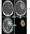
Example of habitat imaging in a patient with glioblastoma multiforme. Habitat imaging (lower right) is obtained by the combination of contrast-enhanced T1W, T2W, and fluid-attenuated inversion recovery (FLAIR) images. Each voxel of a tumour is assigned a specific colour depending on the combination of signal intensity (high/low) of these sequences, e.g., the red voxel is low on T1W images, and high on T2W and FLAIR images in this case. The clusters of voxels with specific colours yield regions that reflect different physiologic microenvironments, called habitats. This regional analysis would help the deeper understanding of tumour heterogeneity. Figure provided courtesy of R. A. Getenby.

References

    1. de Bruin EC, McGranahan N, Mitter R, et al. Spatial and temporal diversity in genomic instability processes defines lung cancer evolution. Science (New York, NY) 2014;346(6206):251–6. - PMC - PubMed
    1. Yates LR, Gerstung M, Knappskog S, et al. Subclonal diversification of primary breast cancer revealed by multiregion sequencing. Nat Med. 2015;21(7):751–9. - PMC - PubMed
    1. Schwarz RF, Ng CK, Cooke SL, et al. Spatial and temporal heterogeneity in high-grade serous ovarian cancer: a phylogenetic analysis. PLoS Med. 2015;12(2):e1001789. - PMC - PubMed
    1. Yamamoto S, Maki DD, Korn RL, Kuo MD. Radiogenomic analysis of breast cancer using MRI: a preliminary study to define the landscape. AJR Am J Roentgenol. 2012;199(3):654–63. - PubMed
    1. Wibmer A, Hricak H, Gondo T, et al. Haralick texture analysis of prostate MRI: utility for differentiating non-cancerous prostate from prostate cancer and differentiating prostate cancers with different Gleason scores. Eur Radiol. 2015;25(10):2840–50. - PMC - PubMed

MeSH terms

Substances