Review
. 2016 Oct:43:28-43.
doi: 10.1016/j.yfrne.2016.10.001. Epub 2016 Oct 11.

The neural basis of sex differences in sexual behavior: A quantitative meta-analysis

Affiliations
Review

The neural basis of sex differences in sexual behavior: A quantitative meta-analysis

Timm B Poeppl et al. Front Neuroendocrinol. 2016 Oct.

Abstract

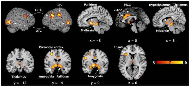
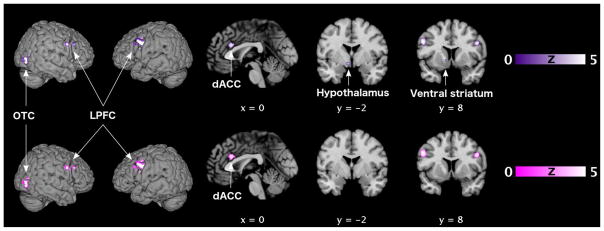
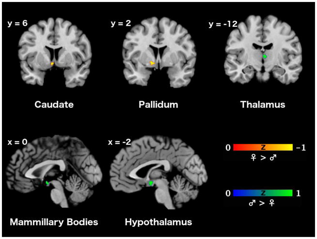
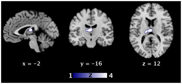
Sexuality as to its etymology presupposes the duality of sexes. Using quantitative neuroimaging meta-analyses, we demonstrate robust sex differences in the neural processing of sexual stimuli in thalamus, hypothalamus, and basal ganglia. In a narrative review, we show how these relate to the well-established sex differences on the behavioral level. More specifically, we describe the neural bases of known poor agreement between self-reported and genital measures of female sexual arousal, of previously proposed male proneness to affective sexual conditioning, as well as hints of unconscious activation of bonding mechanisms during sexual stimulation in women. In summary, our meta-analytic review demonstrates that neurofunctional sex differences during sexual stimulation can account for well-established sex differences in sexual behavior.

Keywords: ALE; Activation likelihood estimation; Functional magnetic resonance imaging; Meta-analysis; Neuroimaging; PET; Positron emission tomography; Sex differences; Sexual behavior; fMRI.


Figures

Figure 1
Figure 1. Functional neuroanatomy of sexual stimulation in women
Significant clusters where the ALE analysis revealed significant convergence of brain activations (p < 0.05, FWE corrected on cluster level) during sexual stimulation in healthy heterosexual women (cf. Table 4). Brain slices are shown at coordinates (x, y, z) in MNI space. ALE, activation likelihood estimation; dACC, dorsal anterior cingulate cortex; FWE, familywise error; IPL, inferior parietal lobule; LPFC, lateral prefrontal cortex; MCC, middle cingulate cortex; MNI, Montreal Neurological Institute; OTC, occipitotemporal cortex.
Figure 2
Figure 2. Common functional neuroanatomy of sexual stimulation in women and men
Locations of significant convergent brain activity (p < 0.05, FWE corrected on cluster level) in both healthy heterosexual women and men as revealed by conjunction (♀ ∩ ♂) analyses (cf. Tables 5/6). Upper row (violet): modality-independent convergence. Lower row (magenta): convergence for visual sexual stimulation. Brain slices are shown at coordinates (x, y, z) in MNI space. dACC, dorsal anterior cingulate cortex; FWE, familywise error; LPFC, lateral prefrontal cortex; MNI, Montreal Neurological Institute; OTC, occipitotemporal cortex.
Figure 3
Figure 3. Differences in consistency of the functional neuroanatomy of sexual stimulation between women and men
Comparison of brain activity between healthy heterosexual women and men as revealed by subtraction (♂ > ♀, ♀ > ♂) analyses (cf. Tables 5/6). Significance threshold set to p > 0.95 posterior probability, cluster size k ≥ 10 voxels. Upper row: modality-independent differences. Lower row: differences for visual sexual stimulation. Brain slices are shown at coordinates (x, y, z) in MNI space. MNI, Montreal Neurological Institute; OFC, orbitofrontal cortex.
Figure 4
Figure 4. Consistent differences in the functional neuroanatomy of sexual stimulation between women and men
ALE analysis revealed significant convergence of differential brain activations (p < 0.05, FWE corrected on cluster level) during sexual stimulation in the thalamus in healthy heterosexual women and men. Modality-independent analysis and analysis restricted to visual sexual stimulation showed similar results (cf. Tables 5/6). Brain slices are shown at coordinates (x, y, z) in MNI space. ALE, activation likelihood estimation; FWE, familywise error; MNI, Montreal Neurological Institute.

References

    1. Abler B, Kumpfmüller D, Grön G, Walter M, Stingl J, Seeringer A. Neural correlates of erotic stimulation under different levels of female sexual hormones. PLoS One. 2013;8:e54447. - PMC - PubMed
    1. Abler B, Seeringer A, Hartmann A, Grön G, Metzger C, Walter M, Stingl J. Neural correlates of antidepressant-related sexual dysfunction: A placebo-controlled fMRI study on healthy males under subchronic paroxetine and bupropion. Neuropsychopharmacology. 2011;36:1837–47. - PMC - PubMed
    1. Acevedo BP, Aron A, Fisher HE, Brown LL. Neural correlates of long-term intense romantic love. Soc Cogn Affect Neurosci. 2012;7:145–59. - PMC - PubMed
    1. Alexander GE, Crutcher MD. Functional architecture of basal ganglia circuits: Neural substrates of parallel processing. Trends Neurosci. 1990;13:266–71. - PubMed
    1. Alekseyenko OV, Waters P, Zhou H, Baum MJ. Bilateral damage to the sexually dimorphic medial preoptic area/anterior hypothalamus of male ferrets causes a female-typical preference for and a hypothalamic Fos response to male body odors. Physiol Behav. 2007;90:438–49. - PMC - PubMed

LinkOut - more resources