. 2016 Nov 15;349(1):119-127.
doi: 10.1016/j.yexcr.2016.10.005. Epub 2016 Oct 11.

Claudin-1 promotes TNF-α-induced epithelial-mesenchymal transition and migration in colorectal adenocarcinoma cells

Affiliations

Claudin-1 promotes TNF-α-induced epithelial-mesenchymal transition and migration in colorectal adenocarcinoma cells

Ajaz A Bhat et al. Exp Cell Res. .

Abstract

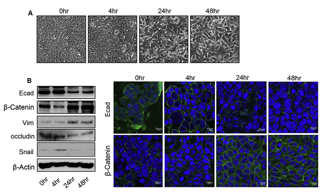
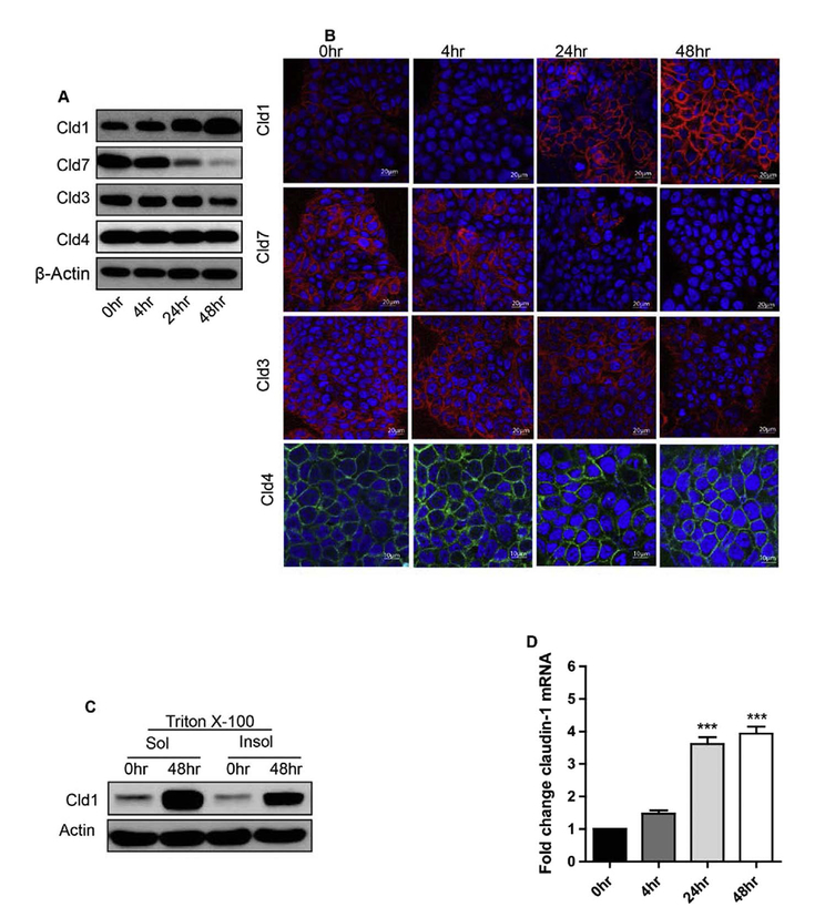
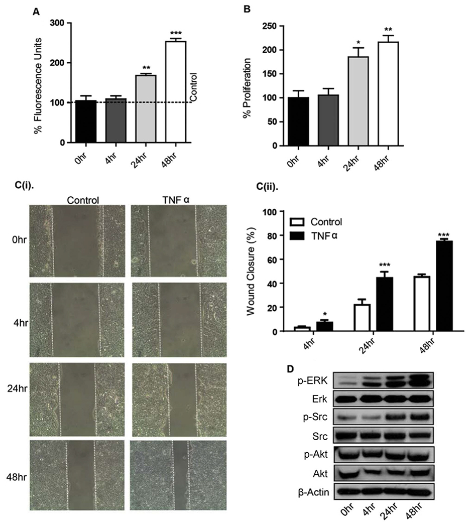
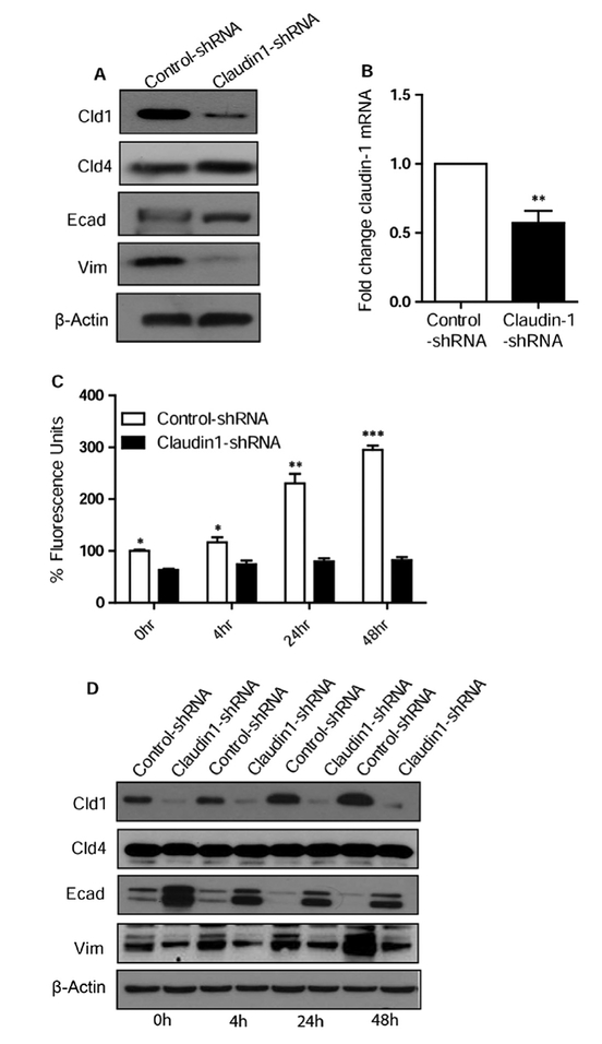
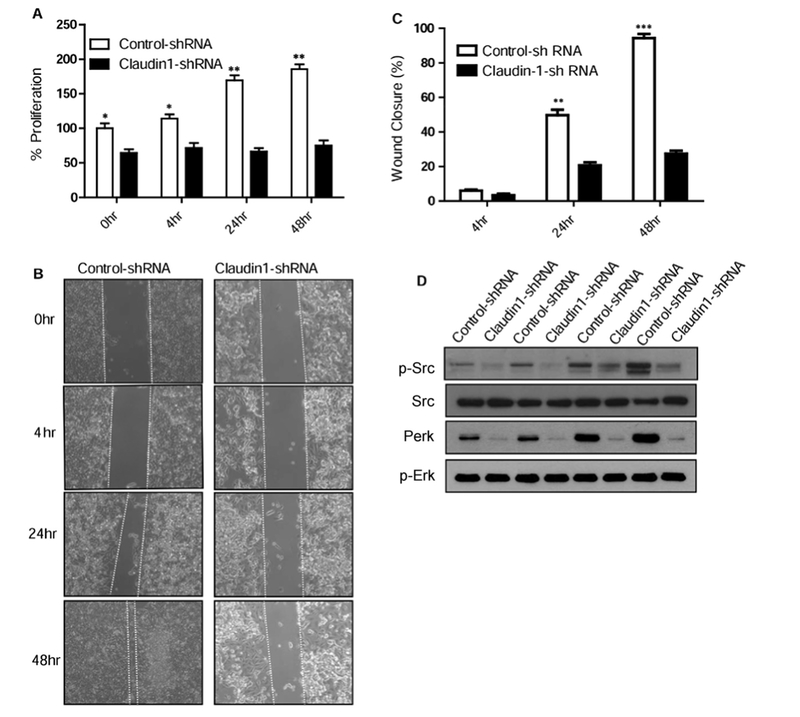
Epithelial-mesenchymal transition (EMT) is an important mechanism in cancer progression and malignancy including colorectal cancer (CRC). Importantly, inflammatory mediators are critical constituents of the local tumor environment and an intimate link between CRC progression and inflammation is now validated. We and others have reported key role of the deregulated claudin-1 expression in colon carcinogenesis including colitis-associated colon cancer (CAC). However, the causal association between claudin-1 expression and inflammation-induced colon cancer progression remains unclear. Here we demonstrate, TNF-α, a pro-inflammatory cytokine, regulates claudin-1 to modulate epithelial to mesenchymal transition (EMT) and migration in colon adenocarcinoma cells. Importantly, colon cancer cells cultured in the presence of TNF-α (10ng/ml), demonstrated a sharp decrease in E-cadherin expression and an increase in vimentin expression (versus control cells). Interestingly, TNF-α treatment also upregulated (and delocalized) claudin-1 expression in a time-dependent manner accompanied by increase in proliferation and wound healing. Furthermore, similar to our previous observation that claudin-1 overexpression in CRC cells induces ERK1/2 and Src- activation, signaling associated with colon cancer cell survival and transformation, TNF-α-treatment induced upregulation of phospho-ERK1/2 and -Src expression. The shRNA-mediated inhibition of claudin-1 expression largely abrogated the TNF-α-induced changes in EMT, proliferation, migration, p-Erk and p-Src expression. Taken together, our data demonstrate TNF-α mediated regulation of claudin-1 and tumorigenic abilities of colon cancer cells and highlights a key role of deregulated claudin-1 expression in inflammation-induced colorectal cancer growth and progression, through the regulation of the ERK and Src-signaling.

Keywords: Claudin-1; EMT; Migration; TNF-alpha.


Figures

Fig. 1.
Fig. 1.
TNFα induces epithelial to mesenchymal transition (EMT) in human colon adenocarcinoma HT29 cells. A. Representative phase-contrast images of HT29 cells growing in monolayer cultures. Treatment of TNFα (10 ng/ml) induced morphological alterations characterized as fibroblast-like cells with significant changes at 24 and 48 h. B. Immunoblot analysis of HT29 cells treated with TNFα for 0–48 h. Cells were lysed in RIPA Buffer, and a total of 20 μg proteins for each sample were loaded onto the SDS-polyacrylamide gel. Membranes were blotted against E-cadherin, β –catenin, vimentin, and occludin whereas actin was used as a loading control. C. Immunofluorescence illustration for localization of E-cadherin and β-catenin with DAPI in HT29 cells treated with TNFα for time dependent manner (0–48 h).
Fig. 2.
Fig. 2.
TNFα induces increase in claudin-1 expression and also its delocalization in human colon adenocarcinoma HT29 cells. A. Immunoblot analysis of HT29 cells treated with TNFα were immune-blotted against claudin-1, claudin-7, claudin-3 and claudin-4. Actin was used as a loading control. B. Immunofluorescence microscopy was performed to analyze the localization of claudin-1, claudin-7, claudin-3 and claudin-4 with DAPI in HT29 cells treated with TNFα for 0–48 h. C. Claudin-1 expression increased in both Tritox-100 soluble and insoluble fractions during the time course of TNFα in HT-29 cells. D. Quantitative Real-Time PCR analysis. HT29 cells were treated with TNFα for 0–48 h, and quantitative RT-PCR was performed. After treatment, total RNA was isolated and subjected to real-time PCR using claudin-1 gene specific primers. Results were plotted as mean ± SD from three independent experiments and presented as fold change. ***p < 0.001 when compared with 24 h and 48 h time points.
Fig. 3.
Fig. 3.
TNF-α promotes permeability, proliferation, and migration of HT29 cells. A. TNFα caused a time dependent increase in paracellular permeability for FITC-dextran (4 kDa). Medium containing FITC-dextran (4 kDa) was added to the top (inner) chamber of the transwell and samples were collected from the bottom chamber after 0, 4, 24 and 48 h after TNFα treatment. B. Cellular proliferation was measured in HT29 cells treated with TNFα (10 ng/ml) for 0–48 h using MTS reagent after plating equal number of cells. C(i). TNF-α enhanced cell migration in a wound healing assay. After mechanical wounding, confluent HT29 cells were pretreated with mitomycin O/N and then treated with TNFα (10 ng/ml) for 0–48 h. Representative photomicrographs of the wounded cell monolayer are shown. C(ii). Percentage of wound closure at 4, 24 and 48 h was calculated. D. Effects of TNF-α on phosphorylation of Erk, Src, and Akt. HT29 cells, which had been serum starved for 1 d, were treated with 10 ng/ml TNFα for the indicated times. Western immunoblots of cellular extracts separated on SDS-polyacrylamide gels were probed with antibodies specific for P-Erk, P-Src, and, P-Akt and these blots were re-probed with respective total Erk, Src, and Akt antibodies. Results were plotted as mean ± SD from three independent experiments and presented as percent change. *p < 0.05**p < 0.01, ***p < 0.001 were considered significant change when compared with control.
Fig. 4.
Fig. 4.
Loss of claudin-1 induces the mesenchymal to epithelial transition (MET) in HT-29 cells. A. Immunoblot analysis of HT29 control and claudin-1 shRNA transfected cells. Cells were lysed in RIPA Buffer, and a total of 20 μg proteins for each sample were loaded onto the SDS-polyacrylamide gel. Membranes were blotted against claudin-1, claudin-4, E-cadherin, and vimentin. Actin was used as a loading control. B. Quantitative Real-Time PCR analysis of claudin-1 in HT-29 cells, transfected with control and claudin-1 shRNA. C. FITC dextran permeability remained significantly less during the time course in claudin-1 knockdown cells compared to control. D. Immuno-blot analysis showed that Claudin-1 knockdown induces MET with time and abrogates the TNF-α mediated changes. Results were plotted as mean ± SD from three independent experiments and presented as percent change. *p < 0.05**p < 0.01, ***p < 0.001 were considered signifi-cant change when compared with control.
Fig. 5.
Fig. 5.
Claudin-1 knockdown reduced TNFα -induced changes in proliferation, Migration, EMT markers and Erk/Src signaling in HT29 cells. A. Cellular proliferation was measured in control or Claudin-1 shRNA transfected HT29 cells treated with or without TNFα (10 ng/ml) using MTS reagent after plating equal number of cells. B. Claudin-1 expression levels affect cell migration. Down regulation of Claudin-1 with shRNA reduced TNFα enhanced migration of HT29 cells. The control or Claudin-1 shRNA transfected HT29 cells were cultured until confluent, mechanically wounded, and then treated with or without 10 ng/ml TNFα. Representative photomicrographs of wounded cell monolayer are shown. C. Percentage of wound closure in each condition was calculated. D. Immunoblot analysis of HT29 control and claudin-1 shRNA transfected cells treated with TNFα for 0–48 h. Cells were lysed in RIPA Buffer, and a total of 20 μg proteins for each sample were loaded onto the SDS-polyacrylamide gel. Membranes were blotted against P-Erk, P-Src, and antibodies against total Erk and Src. Results were plotted as mean ± SD from three independent experiments and presented as percent change. *p < 0.05**p < 0.01, ***p < 0.001 were considered significant change when compared control.

References

    1. Dhawan P, Singh AB, Deane NG, No Y, Shiou SR, Schmidt C, et al., Claudin-1 regulates cellular transformation and metastatic behavior in colon cancer, J. Clin. Investig 115 (7) (2005) 1765–1776. - PMC - PubMed
    1. Kinugasa T, Akagi Y, Yoshida T, Ryu Y, Shiratuchi I, Ishibashi N, et al., Increased claudin-1 protein expression contributes to tumorigenesis in ulcerative colitis-associated colorectal cancer, Anticancer Res 30 (8) (2010) 3181–3186. - PubMed
    1. Suren D, Yildirim M, Kaya V, Alikanoglu AS, Bulbuller N, Yildiz M, et al., Loss of tight junction proteins (Claudin 1, 4, and 7) correlates with aggressive behavior in colorectal carcinoma, Med. Sci. Monit 20 (2014) 1255–1262. - PMC - PubMed
    1. Pope JL, Ahmad R, Bhat AA, Washington MK, Singh AB, Dhawan P, Claudin-1 overexpression in intestinal epithelial cells enhances susceptibility to adenamatous polyposis coli-mediated colon tumorigenesis, Mol. Cancer 13 (2014) 167. - PMC - PubMed
    1. Nakagawa S, Miyoshi N, Ishii H, Mimori K, Tanaka F, Sekimoto M, et al., Expression of CLDN1 in colorectal cancer: a novel marker for prognosis, Int. J. Oncol 39 (4) (2011) 791–796. - PubMed

MeSH terms