. 2016 Dec 15;76(24):7208-7218.
doi: 10.1158/0008-5472.CAN-16-1681. Epub 2016 Oct 14.

A Hyaluronidase-Responsive Nanoparticle-Based Drug Delivery System for Targeting Colon Cancer Cells

Affiliations

A Hyaluronidase-Responsive Nanoparticle-Based Drug Delivery System for Targeting Colon Cancer Cells

Mingzhen Zhang et al. Cancer Res. .

Abstract

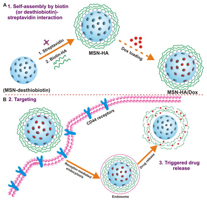
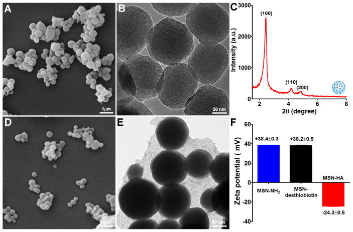
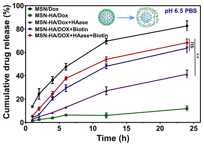
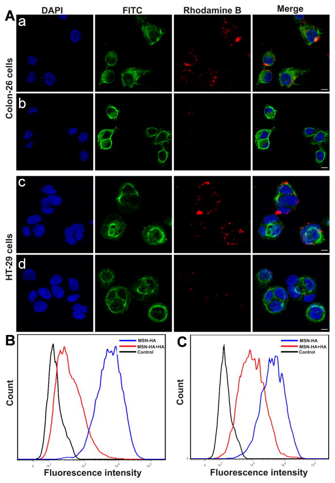
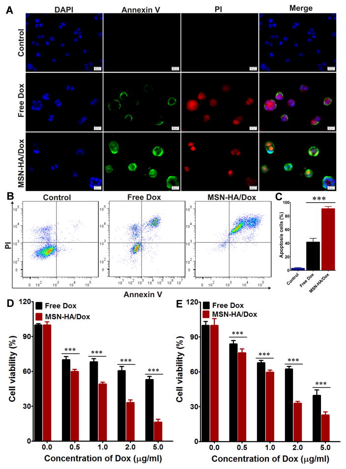
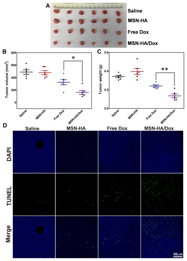
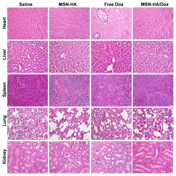
The ability of nanoparticles to target tumors and to enable site-specific drug release provides a unique system for the delivery of effective therapy with reduced toxic side effects. In this study, we used mesoporous silica nanoparticles (MSN) to fabricate a targeted drug delivery system that is responsive to hyaluronidase (HAase). Following engraftment of desthiobiotin onto the surface of MSN, a streptavidin complex was generated, which was functionalized with biotin-modified hyaluronic acid (HA) to enable controlled drug release at cancer cells expressing HAase. Various technologies were used to confirm the successful fabrication of this MSN-based nanocarrier system for targeted drug delivery. In vitro analyses showed that the release of doxorubicin hydrochloride (Dox) was accelerated significantly in the presence of biotin or HAase and accelerated further in the presence of biotin and HAase. Uptake by cancer cells was mediated efficiently by CD44 receptor-mediated endocytosis and the MSN exhibited good biocompatibility in vitro and in vivo MSN-HA/Dox nanoparticles induced apoptosis in cancer cells more efficiently than free doxorubicin and inhibited tumor growth with minimal systemic toxicity in vivo Collectively, our findings offered a preclinical proof of concept for a novel targeted drug delivery carrier system for cancer therapy. Cancer Res; 76(24); 7208-18. ©2016 AACR.


Conflict of interest statement

Confliction of Interest: The authors declare no conflict of interest.

Figures

Figure 1
Figure 1. Schematic diagram to describe MSN-HA nanoparticles mediated delivery of the therapeutic drug, doxorubicin (Dox), to cancer cells
A. Drug loading steps to yield MSN-HA/Dox delivery system. Propylamine functionalized silica (MSN-NH2) was first modified with desthiobiotin to obtain MSN-desthiobiotin, then by employing biotin (or desthiobiotin)-SA interaction, SA and biotinylated HA were self-assembled on the external surface of MSN to yield MSN-HA. Optionally, therapeutic drug, Dox, was able to load to obtain MSN-HA/Dox. B. Schematic illustration of the CD44-receptor-mediated endocytosis and triggering of drug release in tumor cells. MSN-HA/Dox were taken up by cancer cell via receptor-mediated endocytosis (HA-CD44 interaction), then loaded Dox was release from the pore of MSN by the triggering of HAase and intracellular biotin.
Figure 2
Figure 2. Characterization of MSN-NH2 and MSN-HA nanoparticles
A. SEM images were performed to characterize MSN-NH2 nanoparticles, scale bar: 1 μm. B. TEM images were performed to characterize MSN-NH2 nanoparticles, scale bar: 50 nm. C. Small-angle power X-ray diffraction was employed to characterize the structure of MSN-NH2. The high ordered lattice array indicated MSN-NH2 has a uniform and well-defined mesostructure. D. SEM images of MSN-HA, scale bar: 1 μm. E. TEM images of MSN-HA, scale bar: 50 nm. F. Zeta potentials of MSN-NH2, MSN-desthiobiotin, and MSN-HA were measured. MSN-NH2 and MSN-desthiobiotin showed a positive surface charge, after grafting HA, surface charge was changed to negative, indicating the successful link of HA, (n=3).
Figure 3
Figure 3
The biotin- and HAase-responsive release profiles of Dox were evaluated. Drug release under pH 6.5 was conducted to mimic the condition of tumor microenvironment. Under different stimulus condition, biotin (2 μM), HAase (150U/ml) or both were added to MSN-HA/Dox solution; as a control, MSN-Dox was employed. At specified time points (1, 2, 4, 6, 12, and 24 h), cumulative drug release were measured and compared, (n=3).
Figure 4
Figure 4
Endocytosis pathway of MSN-HA taken up by Colon-26 and HT-26 cells was investigated. A. Confocal microscopic images show: a, Colon-26 cells treated with MSN-HA in the absence of HA; b, Colon-26 cells treated with MSN-HA in the presence of HA (2mg/ml); c, HT-29 cells treated with MSN-HA in the absence of HA; d, HT-29 cells treated with MSN-HA in the presence of HA (2mg/ml). Blue channel: DAPI; Green channel: FITC; Red channel: the fluorescence of rhodamine B. The fluorescence intensities of rhodamine B-labeled MSN-HA applied with or without HA were quantified by flow cytometry. B. Colon-26 cells. C. HT-29 cells. (n = 5). Scale bar: 20 μm.
Figure 5
Figure 5
Evaluation the apoptosis induced by MSN-HA/Dox in vitro. A. Fluorescence imaging of Colon-26 cells treated with free Dox or MSN-HA/Dox for 8 h and then co-stained with Annexin V-FTIC and PI. Scale bar: 20 μm. B. Flow cytometric analysis of apoptosis in Colon-26 cells treated with free Dox or MSN-HA/Dox for 8 h. C. Quantification of the Annexin V-FTIC/PI-positive apoptotic cells from panel B. Data are from three independent experiments. D. The apoptotic effects of free Dox and MSN-HA/Dox in Colon-26 cells were assessed by MTT assay, (n=5). E. The apoptotic effects of free Dox and MSN-HA/Dox in HT-29 cells were assessed by MTT assay, (n=5), ***p < 0.001.
Figure 6
Figure 6
Effects of MSN-HA/Dox against Colon-26 xenograft tumors were evaluated in vivo. A. Representative Photos of tumor tissues obtained from tumor-bearing mice treated for 18 days with saline (control), MSN-HA, free Dox, or MSN-HA/Dox, (n=6). B. Tumor volumes were measured at the end of the experiment (n=6). C. Tumor weights were measured at the end of the experiment (n=6). D. TUNEL staining was used to examine apoptosis in tumor sections (green, TUNEL positive cells; blue, cell nuclei), (n=4). Scale bar: 50 μm; *p < 0.05 and **p < 0.01.
Figure 7
Figure 7
The major organs of tumor-bearing mice treated with saline, MSN-HA, free Dox, or MSN-HA/Dox were subjected to histological examination. Heart samples of free Dox-treated mice show intensive vacuolization and myofibril loss (as indicated). Scale bar: 20 μm.

References

    1. Thun MJ, DeLancey JO, Center MM, Jemal A, Ward EM. The global burden of cancer: priorities for prevention. Carcinogenesis. 2010;31:100–10. - PMC - PubMed
    1. Brigger I, Dubernet C, Couvreur P. Nanoparticles in cancer therapy and diagnosis. Adv Drug Deliv Rev. 2012;64:24–36. - PubMed
    1. Xiao B, Zhang M, Viennois E, Zhang Y, Wei N, Baker MT, et al. Inhibition of MDR1 gene expression and enhancing cellular uptake for effective colon cancer treatment using dual-surface-functionalized nanoparticles. Biomaterials. 2015;48:147–60. - PMC - PubMed
    1. Wei T, Liu J, Ma H, Cheng Q, Huang Y, Zhao J, et al. Functionalized nanoscale micelles improve drug delivery for cancer therapy in vitro and in vivo. Nano Lett. 2013;13:2528–34. - PubMed
    1. Kanamala M, Wilson WR, Yang M, Palmer BD, Wu Z. Mechanisms and biomaterials in pH-responsive tumour targeted drug delivery: A review. Biomaterials. 2016;85:152–67. - PubMed

Publication types