. 2016 Nov 1;113(44):E6831-E6839.
doi: 10.1073/pnas.1603529113. Epub 2016 Oct 14.

Neural tube morphogenesis in synthetic 3D microenvironments

Affiliations

Neural tube morphogenesis in synthetic 3D microenvironments

Adrian Ranga et al. Proc Natl Acad Sci U S A. .

Erratum in

Abstract

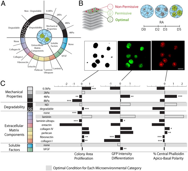
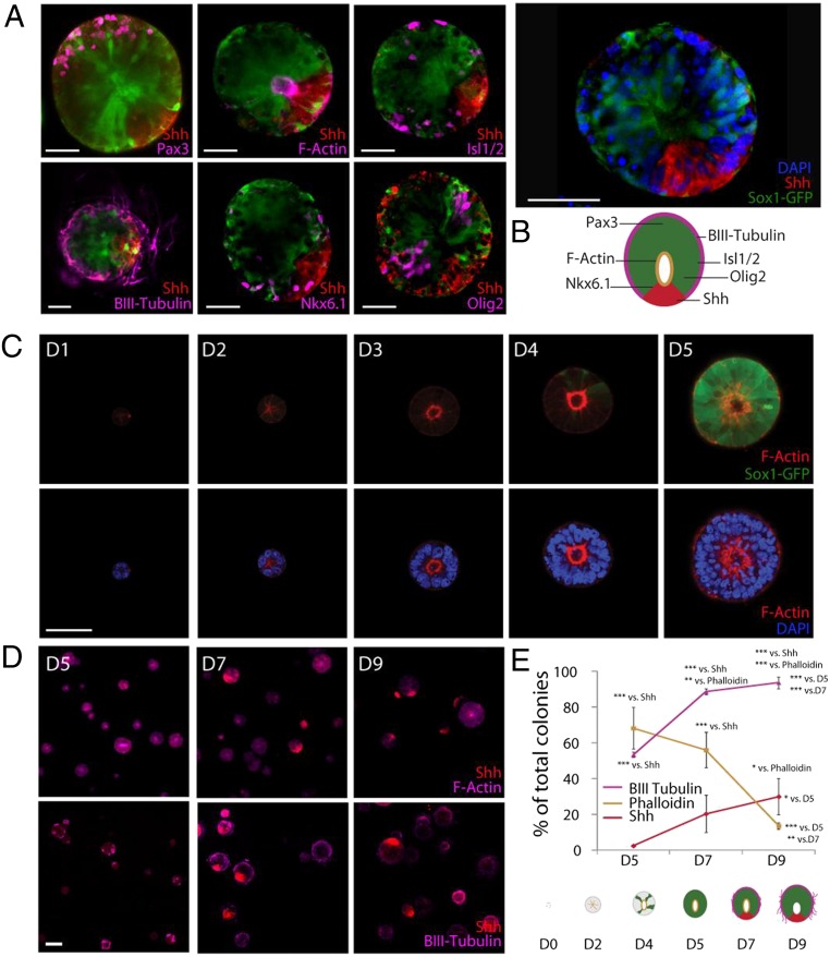
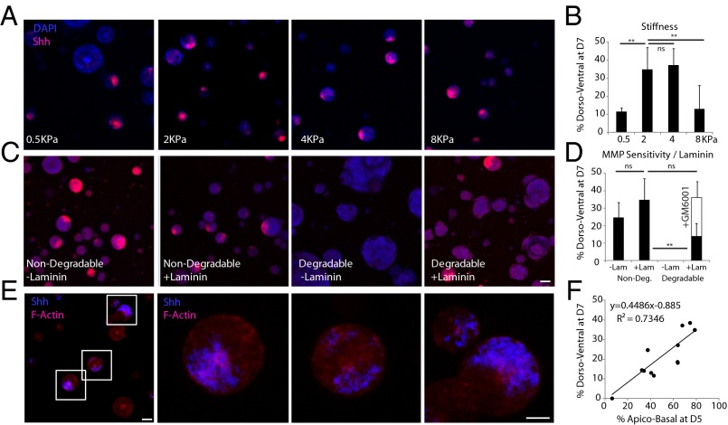
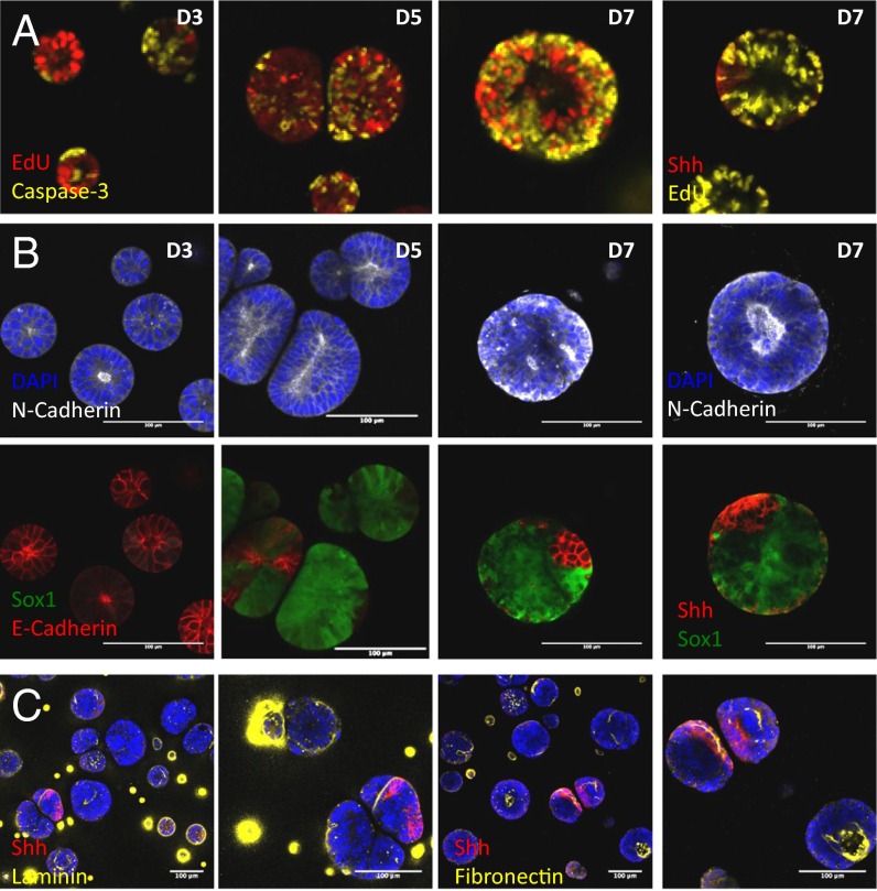
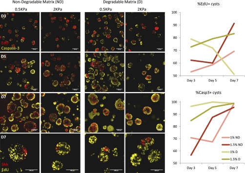
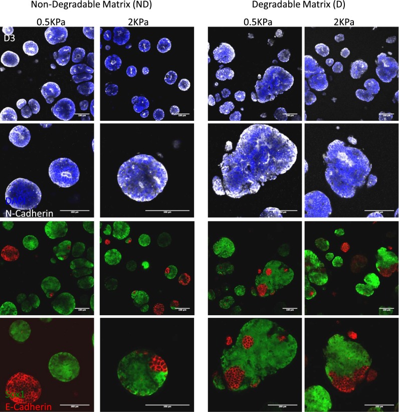
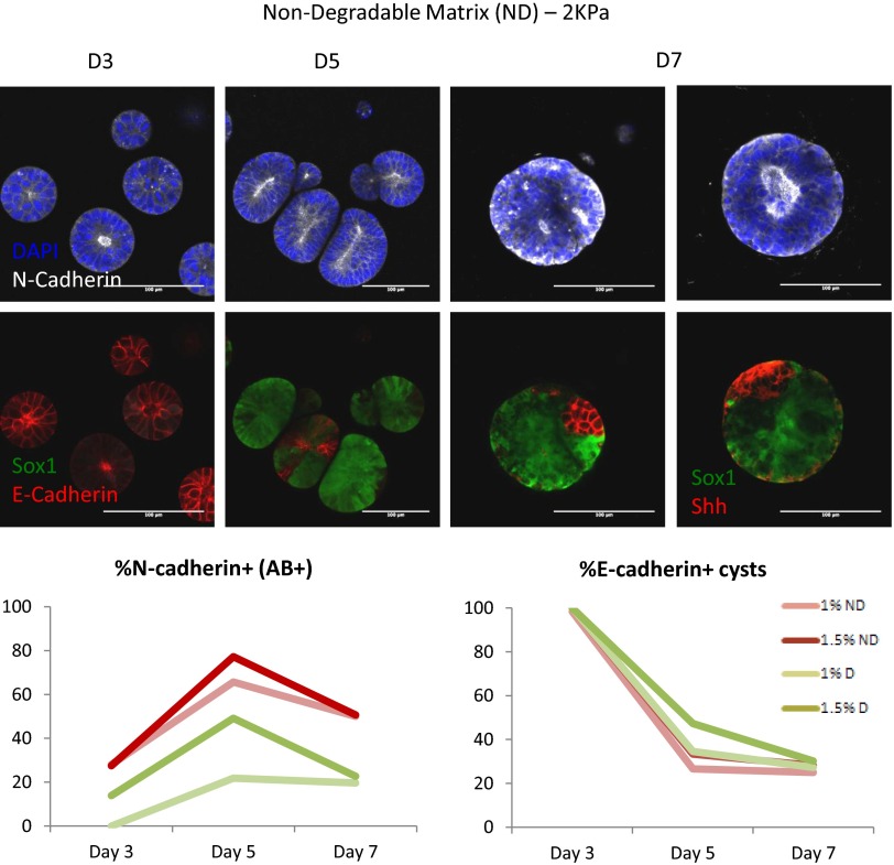
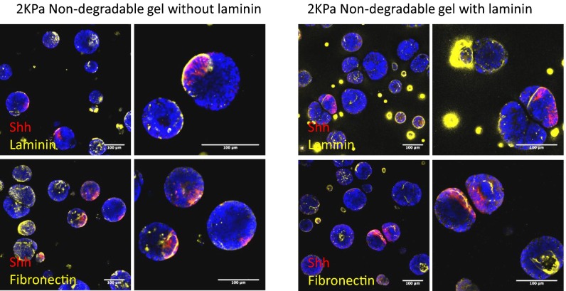
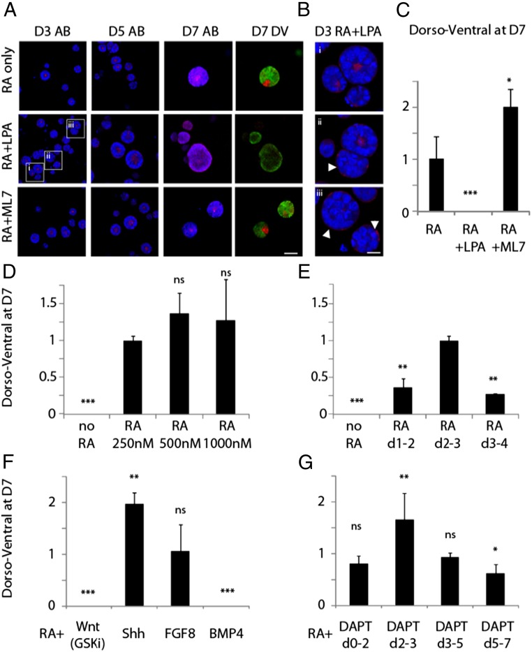
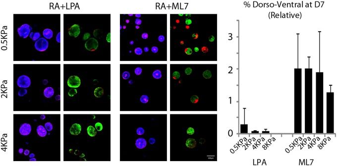
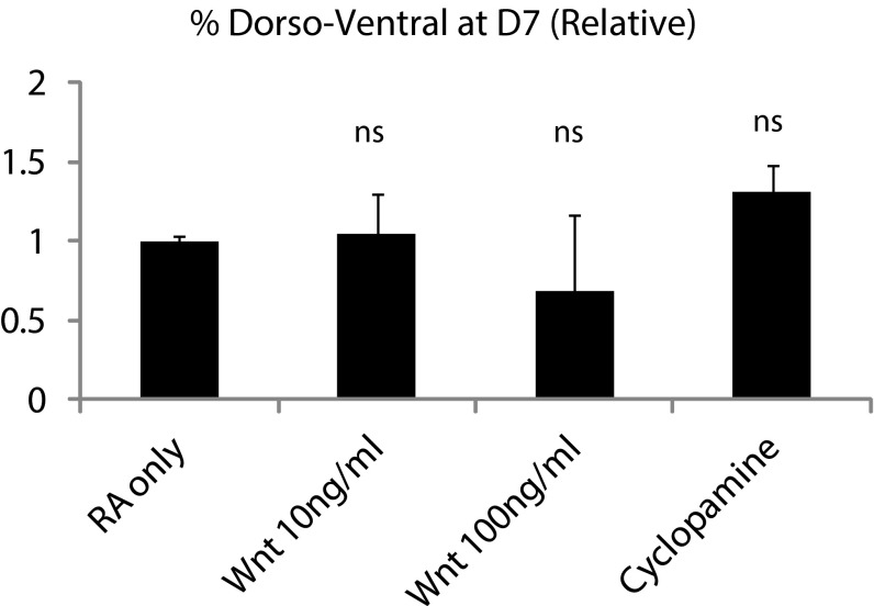
Three-dimensional organoid constructs serve as increasingly widespread in vitro models for development and disease modeling. Current approaches to recreate morphogenetic processes in vitro rely on poorly controllable and ill-defined matrices, thereby largely overlooking the contribution of biochemical and biophysical extracellular matrix (ECM) factors in promoting multicellular growth and reorganization. Here, we show how defined synthetic matrices can be used to explore the role of the ECM in the development of complex 3D neuroepithelial cysts that recapitulate key steps in early neurogenesis. We demonstrate how key ECM parameters are involved in specifying cytoskeleton-mediated symmetry-breaking events that ultimately lead to neural tube-like patterning along the dorsal-ventral (DV) axis. Such synthetic materials serve as valuable tools for studying the discrete action of extrinsic factors in organogenesis, and allow for the discovery of relationships between cytoskeletal mechanobiology and morphogenesis.

Keywords: development; embryonic stem cell; extracellular matrix; neural tube; organoid.


Conflict of interest statement

The authors declare no conflict of interest.

Figures

Fig. 1.
Fig. 1.
A library of molecular building blocks are mixed and cross-linked in situ to form cell-containing 3D hydrogels with independently controllable mechanical and biochemical properties (A). Colonies are differentiated over 5 d, with RA treatment at D2–3. A Sox1-GFP ESC reporter line is used to visualize neural differentiation. Apical–basal polarity is evaluated based on the presence of a characteristic actomyosin contractile ring upon phalloidin staining of the actin cytoskeleton. These markers, along with colony size as an indicator of proliferation, are assessed using an automated imaging and image analysis pipeline (B). (Scale bar: 50 µm.) Systematic investigation of the role of the microenvironment is performed by GLMs, yielding the relative contribution of each factor; statistical analysis identifies the significance of the change with respect to the condition in white for every category (C). Data are shown normalized and centered across the experiment, where a result of 0 indicates a value that corresponds to the mean for the entire experiment. A “signature of differentiation” consisting of a compromise optimal condition within each category across the readouts (proliferation, differentiation, and apical–basal polarity) is thus identified and highlighted in gray.
Fig. S1.
Fig. S1.
Multidimensional analysis of high-throughput dataset.
Fig. S2.
Fig. S2.
Clustering analysis relating multiple inputs to corresponding microenvironments. Clustered output metrics for each well in the screen, with representative clusters identified and further described in terms of microenvironmental contributions. Solidity is defined as the ratio of between the colony area and the convex hull enclosing it. Eccentricity is the ratio between the minor and major axes of an ellipse enclosing the colony. Both measures are equal to 1 for a perfectly round colony.
Fig. 2.
Fig. 2.
Neuroepithelial cysts in Matrigel and optimal PEG hydrogel condition at D2 (A and B, respectively) and D5 (C and D), with additional representative colonies at D5 (E and F). Colony area was quantified for both conditions within a representative time series (G), with a comparison of ensuing apical–basal polarity evaluated at D5 (H). (Scale bars: 50 µm.) **P < 0.01.
Fig. 3.
Fig. 3.
Cyst identity is characterized by markers of DV patterning in optimal synthetic matrix conditions (A). Cyst patterning exhibits key features of neural tube architecture, including floor plate (Shh), progenitor motor neuron (Olig2), motor neuron (Isl1/2), postmitotic neuron (βIII-tubulin), as well as ventral (Nkx6.1) and dorsal (Pax3) positional identities, represented schematically (B). Representative images of a time series with Sox1-GFP expression and F-Actin staining (D1 to D5) (C), and with Shh, F-Actin, and βIII-tubulin staining (D5 to D9) (D). Quantification of key events in morphogenetic patterning in 3D (E). (Scale bars: A and C, 50 µm; D, 100 µm.) ***P < 0.001, **P < 0.01, *P < 0.05.
Fig. 4.
Fig. 4.
Representative images of Shh-stained cysts in nondegradable matrices of various stiffness (A) and in 2-kPa matrices of various degradability and laminin compositions (B) with corresponding quantification of DV patterning efficiency for each condition (C and D). F-actin and Shh costaining of cysts in nondegradable, 2-kPa, laminin-containing matrix, indicating apical–basal and DV patterning, respectively, with higher magnification images (E). The relationship between apical–basal polarity at D5 and DV patterning at D7 is quantified for all tested conditions (F). [Scale bars: A, C, and E (Left), 100 µm; E (Right), 50 µm.] **P < 0.01.
Fig. 5.
Fig. 5.
Characterization of proliferation/apoptosis, neural/epithelial identity, and ECM production in standard 2KPa MMP-insensitive synthetic matrix. (A) Cysts are highly proliferative at D3, followed by gradual apoptosis mainly at the basal aspect. Proliferation is absent in the Shh region. (B) N-Cadherin is apically-basally polarized in cysts which undergo morphogenesis. E-Cadherin is present in all cysts at D3 and is rapidly lost by D5. E-Cad is still present in some cysts up to D7. (C) Cysts produce laminin and fibronectin, frequently localized at the Shh polarized and apical region of the cyst. (Scale bar: 100 μm.)
Fig. S3.
Fig. S3.
Characterization and dynamics of proliferation and apoptosis during 3D differentiation.
Fig. S4.
Fig. S4.
Characterization of neural and epithelial markers at D7 of 3D differentiation.
Fig. S5.
Fig. S5.
Dynamics of neural and epithelial markers during 3D differentiation. AB+, Apico–basally polarized.
Fig. S6.
Fig. S6.
ECM markers at D7 in laminin and laminin-free synthetic matrix.
Fig. 6.
Fig. 6.
Comparison of the effect of LPA and ML7 treatments on cyst apical–basal (AB) polarity at D3, D5, and D7, and on dorsal–ventral (DV) patterning at D7 (A), with corresponding quantification (C). High-magnification images of mixed polarity and inversely polarized cysts after LPA treatment at D3 (B). Effects of modulating RA concentration at D2–3 (D), modulating RA treatment time (at standard RA concentration) (E), developmental signaling pathway modulation at D2–3 (F), and Notch inhibition at various time points (G). All experiments in standard optimal matrix conditions (nondegradable, 2 kPa, with laminin), percentage DV efficiency corresponding to standard RA treatment (D2–3, 250 nM) set at 1, and all other treatments normalized to that of standard RA treatment (dorsoventral at D7 = % DV for treatment/% DV for standard). (Scale bars: A, 100 µm; B, 50 µm.) ***P < 0.001, **P < 0.01, *P < 0.05.
Fig. S7.
Fig. S7.
Activation and inhibition of the RhoA/ROCK pathway over multiple stiffness range.
Fig. S8.
Fig. S8.
Effect of Wnt3a protein and cyclopamine on DV patterning.

References

    1. Kojima Y, Tam OH, Tam PP. Timing of developmental events in the early mouse embryo. Semin Cell Dev Biol. 2014;34:65–75. - PubMed
    1. Eyckmans J, Boudou T, Yu X, Chen CS. A hitchhiker’s guide to mechanobiology. Dev Cell. 2011;21(1):35–47. - PMC - PubMed
    1. Stern CD, Fraser SE. Tracing the lineage of tracing cell lineages. Nat Cell Biol. 2001;3(9):E216–E218. - PubMed
    1. De Robertis EM. Spemann’s organizer and self-regulation in amphibian embryos. Nat Rev Mol Cell Biol. 2006;7(4):296–302. - PMC - PubMed
    1. Sato T, et al. Single Lgr5 stem cells build crypt-villus structures in vitro without a mesenchymal niche. Nature. 2009;459(7244):262–265. - PubMed

Publication types