Review
. 2017 Feb;35(2):213-223.
doi: 10.1002/jor.23460. Epub 2016 Dec 19.

Fracture healing physiology and the quest for therapies for delayed healing and nonunion

Affiliations
Review

Fracture healing physiology and the quest for therapies for delayed healing and nonunion

Paul Kostenuik et al. J Orthop Res. 2017 Feb.

Abstract

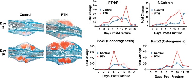
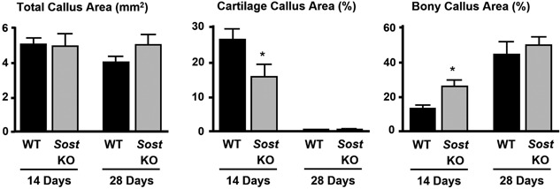
Delayed healing and nonunion of fractures represent enormous burdens to patients and healthcare systems. There are currently no approved pharmacological agents for the treatment of established nonunions, or for the acceleration of fracture healing, and no pharmacological agents are approved for promoting the healing of closed fractures. Yet several pharmacologic agents have the potential to enhance some aspects of fracture healing. In preclinical studies, various agents working across a broad spectrum of molecular pathways can produce larger, denser and stronger fracture calluses. However, untreated control animals in most of these studies also demonstrate robust structural and biomechanical healing, leaving unclear how these interventions might alter the healing of recalcitrant fractures in humans. This review describes the physiology of fracture healing, with a focus on aspects of natural repair that may be pharmacologically augmented to prevent or treat delayed or nonunion fractures (collectively referred to as DNFs). The agents covered in this review include recombinant BMPs, PTH/PTHrP receptor agonists, activators of Wnt/β-catenin signaling, and recombinant FGF-2. Agents from these therapeutic classes have undergone extensive preclinical testing and progressed to clinical fracture healing trials. Each can promote bone formation, which is important for the stability of bridged calluses, and some but not all can also promote cartilage formation, which may be critical for the initial bridging and subsequent stabilization of fractures. Appropriately timed stimulation of chondrogenesis and osteogenesis in the fracture callus may be a more effective approach for preventing or treating DNFs compared with stimulation of osteogenesis alone. © 2016 Orthopaedic Research Society. Published by Wiley Periodicals, Inc. J Orthop Res 35:213-223, 2017.

Keywords: chondrogenesis; delayed union; fracture healing; nonunion fracture; osteogenesis.


Figures

Figure 1
Figure 1
Biomechanical strength recovery of internally stabilized closed femoral fractures created in normal gonad‐intact female rats at 8 weeks of age (white circles), and in OVX rats at 32 (gray circles) or 50 (black circles) weeks of age. Animals were sacrificed at various time points after fracture for biomechanical testing of callus breaking load by a 3‐point bending test. Data represent percentage of the non‐fractured contralateral femur diaphysis, n = 3–10 per group per time point. Fractured femurs from older OVX rats showed slower regain of strength compared with young gonad‐intact rats. Reproduced with permission from Meyer et al.20
Figure 2
Figure 2
Stages of fracture healing in rodents subjected to internally stabilized experimental long bone fractures, as described by Bonnarens and Einhorn.17 Reproduced with permission from Hadjiargyrou et al.111
Figure 3
Figure 3
Impaired long bone fracture healing in PTHrP haplo‐insufficient mice compared with wild‐type (WT) mice. PTHrP(+/−) mice exhibited an early transient deficit in callus cartilage a more sustained deficit in callus size and bone content. Day 7 and 14 refer to the time after creation of internally stabilized closed femoral fracture. Data represent means ± SD, n = 6/group. *Significant differences versus wildtype (WT) controls, p < 0.05. Reproduced with permission from Wang et al.39
Figure 4
Figure 4
Recombinant human BMP2 (rhBMP2) induced cartilage formation during the repair of stabilized closed long bone fractures in mice. Mice received PBS (control) or 10 μg of rhBMP2 by direct injection into the fresh fracture site, and were sacrificed 10 days later. Upper graph: Total callus volume and callus cartilage volume were significantly greater in the rhBMP2 group versus PBS controls when measured by histomorphometry 10 days post‐fracture (**P < 0.05). Lower images: Safranin‐O/Fast Green staining of callus sections, with a higher‐magnification image showing chondrocytes spanning the fracture line that is near the left side of the inset. Reproduced with permission from Yu et al.49
Figure 5
Figure 5
Changes in PTHrP, Wnt signaling (i.e., beta‐catenin), chondrogenesis and osteogenesis in normal mice after creation of internally stabilized closed femur fractures. Mice were treated with PTH(1‐34) (30 μg/kg/day) for 14 days post‐fracture. Healing fractures were harvested at various times and assessed by histology (left panels) or by RNA analyses (right panels). Histology sections obtained 5 and 10 days post‐fracture were stained with Safranin O/fast green, which labels cartilage and chondrogenic cells (red color). The right panels show that PTH(1‐34) increased mRNA expression of PTHrP, the chondrogenic marker Sox9, the osteogenic marker Runx2, and the Wnt signaling marker beta‐catenin. Reproduced with permission from Kakar et al.67
Figure 6
Figure 6
Histomorphometry of callus composition for normal wild‐type (WT) mice and Sost knockout (KO) mice subjected to internally stabilized closed femoral fractures. Data were generated on samples collected 14 or 28 days post‐fracture. Data represent means and SEM, n = 6–10/group/time point. *P < 0.05 for SOST KO versus WT. Reproduced with permission from Li et al.100

References

    1. Mosekilde L, Bak B. 1993. The effects of growth hormone on fracture healing in rats: a histological description. Bone 14:19–27. - PubMed
    1. Tzioupis C, Giannoudis PV. 2007. Prevalence of long‐bone non‐unions. Injury 38:S3–9. - PubMed
    1. Einhorn TA, Gerstenfeld LC. 2015. Fracture healing: mechanisms and interventions. Nat Rev Rheumatol 11:45–54. - PMC - PubMed
    1. Westgeest J, Weber D, Dulai SK, et al. 2016. Factors associated with development of nonunion or delayed healing after an open long bone fracture: a prospective cohort study of 736 subjects. J Orthop Trauma 30:149–155. - PubMed
    1. Marsh D. 1998. Concepts of fracture union, delayed union, and nonunion. Clin Orthop Relat Res S22–30. - PubMed

Publication types