Review
. 2017 Jan;42(1):57-71.
doi: 10.1016/j.tibs.2016.09.004. Epub 2016 Oct 12.

Touch, Tension, and Transduction - The Function and Regulation of Piezo Ion Channels

Affiliations
Review

Touch, Tension, and Transduction - The Function and Regulation of Piezo Ion Channels

Jason Wu et al. Trends Biochem Sci. 2017 Jan.

Abstract

In 2010, two proteins, Piezo1 and Piezo2, were identified as the long-sought molecular carriers of an excitatory mechanically activated current found in many cells. This discovery has opened the floodgates for studying a vast number of mechanotransduction processes. Over the past 6 years, groundbreaking research has identified Piezos as ion channels that sense light touch, proprioception, and vascular blood flow, ruled out roles for Piezos in several other mechanotransduction processes, and revealed the basic structural and functional properties of the channel. Here, we review these findings and discuss the many aspects of Piezo function that remain mysterious, including how Piezos convert a variety of mechanical stimuli into channel activation and subsequent inactivation, and what molecules and mechanisms modulate Piezo function.


Figures

Figure I
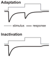
Figure I. Adapting vs. inactivating currents
Idealized currents (black) in response to a two-pulse stimulus protocol (gray), demonstrating the presence of additional current upon an increase in stimulus intensity for an adapting, but not for an inactivating current.
Figure 1
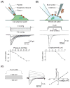
Figure 1. Piezos are mechanically activated ion channels
(A) Schematic of “stretch” setup, in which negative suction is applied to a cell-attached patch with a high-speed pressure clamp through the patch pipette, stimulating only those channels contained within the patch dome (above). Piezo1 peak current amplitudes initially rise with increasing magnitudes of pressure before reaching saturation (middle). The pressure-response relationship can be fit with a sigmoidal function to measure pressure sensitivity (below). Data are from Wu and Grandl, unpublished. (B) Schematic of “poke” setup depicting cell deformation by a blunt probe (typically a fire-polished glass pipette) during a whole-cell recording, which activates a larger population of channels throughout the cell (above). Piezo1 current amplitudes increase with increasing steps of displacement beginning a few micrometers beyond first contact of the probe with the cell membrane. From these experiments, a current-displacement curve can be generated. Typically, currents do not plateau before cell rupture (below). Data are from Lewis and Grandl, unpublished. (C) Voltage step protocol with a single “poke” displacement during each step (left). A family of currents from a single cell illustrates the voltage dependence of channel inactivation, with severely slowed decay times at positive voltages (middle). An I-V curve plotted from peak current amplitudes reveals a reversal potential near 0 mV, demonstrating cationic non-selectivity (right). Data are from Lewis and Grandl, unpublished.
Figure 2
Figure 2. Expression and physiological roles of Piezos
Piezo1 and Piezo2 are expressed in a diverse set of organs and tissues within the human body, contributing to an equally diverse set of physiological roles [, –, , –, , –, , –83]. Numbered tissues are as follows: 1. Brain, 2. Optic nerve head, 3. Periodontal ligament, 4. Trigeminal ganglion, 5. Dorsal root ganglion and skin, 6. Lungs, 7. Cardiovascular system and red blood cells, 8. Gastrointestinal system, 9. Kidney, 10. Colon, 11. Bladder, 12. Articular cartilage. Tissues in which Piezo function has been extensively studied are expanded to show detail. Top left inset illustrates Piezo2 expressed in Merkel cells of the skin, where mechanical activation of Piezo mediates depolarization and activation of dorsal root ganglion cell afferents, which also express Piezo2. Together, these cells are involved in sensing light touch and proprioception. Bottom left inset highlights the expression of both Piezo1 and Piezo2 in chondrocytes of articular cartilage, where they activate under compressive force. Top right inset illustrates the role of Piezo1 in sensing mechanical properties of the environment of neural progenitor cells, thereby initiating signaling pathways that lead to neuronal differentiation and subsequent development of neurite morphology, neuronglia interactions, and nanoroughness of glial membranes. Middle right inset depicts the role of Piezo1 in regulating volume of red blood cells as well as sensing shear stress to regulate vascular branching and alignment of endothelial cells. Bottom right inset shows the role of Piezo1 in sensing fluid flow throughout the nephron of the kidney. Deficits in Piezo1 function in the kidney may lead to downstream effects on urinary osmolarity and renal pathologies.
Figure 3
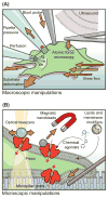
Figure 3. Current and future methods of stimulating Piezos
Orange arrows represent direction of force in relation to cell or channel, and Piezo ion channels are illustrated in red. (A) Macroscopic methods for stimulating large populations of Piezo channels, whose activity can be measured with electrophysiology or through calcium imaging [1, 13, 14, 18]. These include directly deforming the cell with a blunt probe (“poke” assay) or with atomic force microscopy. High pressure perfusion is an alternative method to deform the cell without physically contacting the membrane, while in contrast, shear flow achieved through microfluidic channels applies a parallel stress to the substrate surface. Both positive and negative pressure through a pipette (“stretch” assay) can stimulate single or many Piezo channels. Substrate deformation with flexible membranes and remote vibration of the cell and surrounding milieu through ultrasound are yet untested methods for directly stimulating Piezo channels. (B) Microscopic modes of Piezo stimulation are shown magnified in the context of the plasma membrane [–49, 69]. Deflection of micropillars stimulates single or small populations of Piezo channels through membrane deformation. The agonist Yoda1 directly activates Piezo1, though the mechanism is unknown. Lipids such as cholesterol modulate Piezo function, but have not yet been shown to directly induce activation. In theory, direct activation of the channel could be achieved through magnetic or optical control of nanoparticles bound to specific channel domains; application of force through magnetic nanobeads has been shown to perturb channel function, but neither technique has been shown to directly activate Piezo.
Figure 4
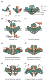
Figure 4. Potential mechanisms of mechanical sensing and activation
(A) The cryo-EM structure (PDB 3JAC) of Piezo1 (left) reveals possible structural domains (right) that may play a role in mechanosensing and channel activation. (B–E) Possible sensing mechanisms and conformational changes by which Piezo channels may activate in response to external forces. Potential ion permeation pathways are indicated with dashed lines; orange represents the closed channel conformation and green represents the open conformation upon applied force (B) Tethering of either the CED domain to the extracellular matrix or the “beams” to cytoskeletal elements may contribute to a gating spring mechanism of activation. (C) Similarly, local shear flow may displace the CED domain and expose an ion permeation pathway. (D) The curved architecture of the cryo-EM Piezo1 structure supports the possibility that Piezo rests in a locally curved lipid bilayer environment. With rising membrane tension, the curvature is reduced, potentially causing hydrophobic mismatch of the “blades” and conformational changes in the “beam” and “anchor” domains to open the pore. (E) Hydrophobic mismatch may also occur due to changes in plasma membrane thickness by in-plane membrane stretch, by which a tilt in the pore helices might lead to pore opening. (F) Annular lipids, agonists, and inhibitors may insert directly within the channel structure to initiate changes in channel conformation. (G) Lipids and chemical modifiers may also insert directly into the membrane causing changes in membrane stiffness, tension, or curvature, leading to channel activation.

References

    1. Coste B, et al. Piezo1 and Piezo2 are essential components of distinct mechanically activated cation channels. Science. 2010;330(6000):55–60. - PMC - PubMed
    1. Kim SE, et al. The role of Drosophila Piezo in mechanical nociception. Nature. 2012;483(7388):209–12. - PMC - PubMed
    1. Coste B, et al. Piezo proteins are pore-forming subunits of mechanically activated channels. Nature. 2012;483(7388):176–81. - PMC - PubMed
    1. Kamajaya A, et al. The structure of a conserved piezo channel domain reveals a topologically distinct beta sandwich fold. Structure. 2014;22(10):1520–7. - PMC - PubMed
    1. Prole DL, Taylor CW. Identification and analysis of putative homologues of mechanosensitive channels in pathogenic protozoa. PLoS One. 2013;8(6):e66068. - PMC - PubMed

Publication types