Review
. 2016 Oct 15;60(2):173-180.
doi: 10.1042/EBC20160005.

Cellular proteostasis: degradation of misfolded proteins by lysosomes

Affiliations
Review

Cellular proteostasis: degradation of misfolded proteins by lysosomes

Matthew P Jackson et al. Essays Biochem. .

Abstract

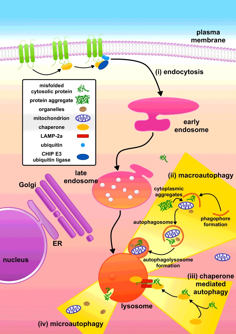
Proteostasis refers to the regulation of the cellular concentration, folding, interactions and localization of each of the proteins that comprise the proteome. One essential element of proteostasis is the disposal of misfolded proteins by the cellular pathways of protein degradation. Lysosomes are an important site for the degradation of misfolded proteins, which are trafficked to this organelle by the pathways of macroautophagy, chaperone-mediated autophagy and endocytosis. Conversely, amyloid diseases represent a failure in proteostasis, in which proteins misfold, forming amyloid deposits that are not degraded effectively by cells. Amyloid may then exacerbate this failure by disrupting autophagy and lysosomal proteolysis. However, targeting the pathways that regulate autophagy and the biogenesis of lysosomes may present approaches that can rescue cells from the deleterious effects of amyloidogenic proteins.

Keywords: Parkinson's disease; TFEB; amyloid; autophagy; chaperone-mediated autophagy; immunoglobulin light chain; lysosome; mTOR; macroautophagy; protein aggregation; protein misfolding; proteostasis; rapamycin; α-synuclein; β2-microglobulin.


Figures

Figure 1
Figure 1. Cellular pathways of protein degradation remove misfolded proteins
Proteins may either fail to fold after their synthesis or fold into their native structure and then subsequently misfold. Cells must dispose of these misfolded proteins and their aggregates in order to maintain proteostasis. Lysosomes and proteasomes represent the principal sites for the degradation of misfolded proteins and play complementary roles in proteostasis.
Figure 2
Figure 2. Multiple pathways deliver proteins to lysosomes for degradation
(i) Misfolded plasma membrane proteins can be tagged for degradation via the E3 ubiquitin ligase CHIP. The resultant ubiquitinated proteins are endocytosed into early endosomes, which mature into late endosomes. The misfolded plasma membrane proteins are then delivered to lysosomes via the fusion of late endosomes with this organelle. (ii) Macroautophagy encapsulates cytoplasmic material, including soluble cytosolic proteins and protein aggregates, into double-membraned vesicles that then fuse with the lysosome. (iii) In CMA, the chaperone Hsc70 recognizes misfolded proteins and these are translocated into the lysosome by LAMP-2a. (iv) Microautophagy is associated with the formation of invaginations in the lysosomal membrane and could also contribute to the delivery of proteins into the lysosome. ER, endoplasmic reticulum. Adapted from [6,9].
Figure 3
Figure 3. Proteins are recognized for CMA by a CMA-targeting motif
(i) The CMA motif may contain up to two hydrophobic residues [isoleucine (I), phenylalanine (F), leucine (L) or valine (V)], up to two positive residues [arginine (R) or lysine (K)] and a single negatively charged residue [glutamate (E) or aspartate (D)] flanked at either the N- or C-terminus of the pentapeptide by a single glutamine (Q) residue. This is often referred to as a KFERQ or KFERQ-like motif [12]. (ii) Protein sequence of α-synuclein highlighting the CMA motif (bold and underlined) and potential CMA-targeting motif residues.

References

    1. Labbadia J., Morimoto R.I. The biology of proteostasis in aging and disease. Annu. Rev. Biochem. 2015;84:435–464. doi: 10.1146/annurev-biochem-060614-033955. - DOI - PMC - PubMed
    1. Gregersen N., Bross P., Vang S., Christensen J.H. Protein misfolding and human Disease. Annu. Rev. Genomics Hum. Genet. 2006;7:103–124. doi: 10.1146/annurev.genom.7.080505.115737. - DOI - PubMed
    1. Ciechanover A. Proteolysis: from the lysosome to ubiquitin and the proteasome. Nat. Rev. Mol. Cell Biol. 2005;6:79–87. doi: 10.1038/nrm1552. - DOI - PubMed
    1. Bhutani N., Piccirillo R., Hourez R., Venkatraman P., Goldberg A.L. Cathepsins L and Z are critical in degrading polyglutamine-containing proteins within lysosomes. J. Biol. Chem. 2012;287:17471–17482. doi: 10.1074/jbc.M112.352781. - DOI - PMC - PubMed
    1. Turk V., Stoka V., Vasiljeva O., Renko M., Sun T., Turk B., Turk D. Cysteine cathepsins: from structure, function and regulation to new frontiers. Biochim. Biophys. Acta. 2012;1824:68–88. doi: 10.1016/j.bbapap.2011.10.002. - DOI - PMC - PubMed

Publication types