

Case Reports
doi: 10.17712/nsj.2016.4.20160280.
Posterior fossa ruptured dermoid cyst presenting with hydrocephalus
Affiliations
- PMID: 27744466
- PMCID: PMC5224435
- DOI: 10.17712/nsj.2016.4.20160280
Item in Clipboard
Case Reports
Posterior fossa ruptured dermoid cyst presenting with hydrocephalus
Neurosciences (Riyadh).
2016 Oct.
Abstract
Dermoid cysts are rare, benign lesions of embryological origin that represent 0.1-0.7% of all intracranial tumors. They are mainly located in the supra tentorial space, especially in the parasellar region. Their location in the posterior fossa remains uncommon. Rupture of intracranial dermoid cysts is a rare phenomenon. We present a case of dermoid cyst, which had ruptured into ventricular system. Computed Tomography and MRI revealed fat in the fourth ventricle, prepontine cistern, and cerebellomedullary cistern. Hydrocephalus was noted. We performed right ventriculo-peritoneal shunt on which patient improved and he continues to remain asymptomatic one year after.
Figures
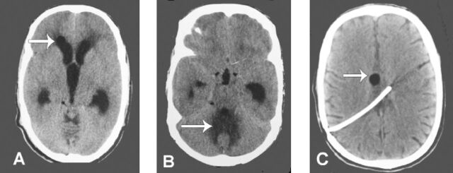
Pre-op non-contrast CT brain a) Features of hydrocephalus with marker showing fat in lateral ventricle, b) Changes in the intensity in vermis, possible site of dermoid cyst after rupture and C) Post-op CT scan decompressed ventricle post–operatively with shunt in situ and marker showing fat.

Pre-op MRI with contrast a) Pre–op (T1 MRI)- Fat in the prepontine, cerebellomedullary cistern and fourth ventricle with hydrocephalus, b) Marked area shows intensity changes in vermis (non enhancement on contrast) possible site of dermoid cyst rupture c) post-op MRI (T1) fat in the same regions with ventricle decompression. Also fat visualised in the lateral ventricle.
References
-
- Benzagmout M, Agharbi S, Chakour K, Chaoui ME. Dermoid cyst of the posterior fossa. Neurosciences (Riyadh) 2011;16:153–155. - PubMed
-
- Liu JK, Gottfried ON, Salzman KL, Schmidt RH, Couldwell WT. Ruptured intracranial dermoid cysts: clinical, radiographic, and surgical features. Neurosurgery. 2008;62:377–384. - PubMed
-
- Cai CQ, Zhang QJ, Hu XL, Wang CX. Dermoid cyst of the posterior fossa associated with congenital dermal sinus in a child. World J Pediatr. 2008;4:66–69. - PubMed
-
- Coulibaly O, Komi E, Rifi L, Sogoba Y, Dama M, Diallo O, et al. Rupture of posterior fossa dermoid cyst overlying the torcular with extracranial extension: technical note. World Journal of Neuroscience. 2015;5:82–86.
Publication types
MeSH terms
LinkOut - more resources
Full Text Sources
Other Literature Sources
Medical