. 2016 Nov 3;99(5):1005-1014.
doi: 10.1016/j.ajhg.2016.08.019. Epub 2016 Oct 13.

Periodontal Ehlers-Danlos Syndrome Is Caused by Mutations in C1R and C1S, which Encode Subcomponents C1r and C1s of Complement

Collaborators, Affiliations

Periodontal Ehlers-Danlos Syndrome Is Caused by Mutations in C1R and C1S, which Encode Subcomponents C1r and C1s of Complement

Ines Kapferer-Seebacher et al. Am J Hum Genet. .

Abstract

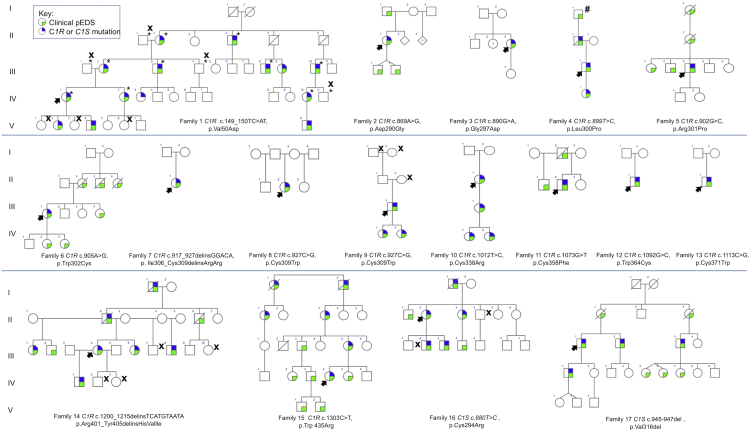
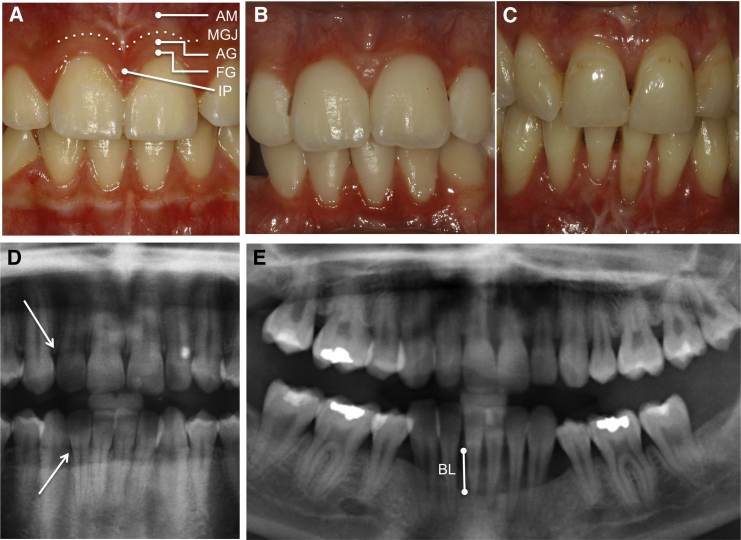
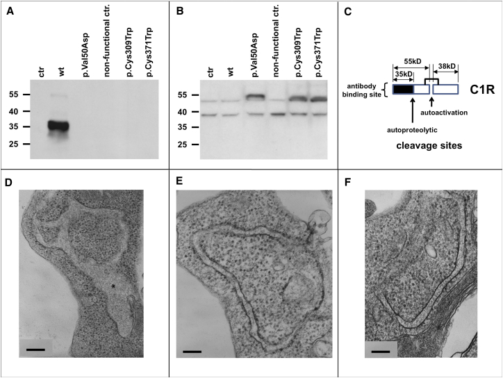
Periodontal Ehlers-Danlos syndrome (pEDS) is an autosomal-dominant disorder characterized by early-onset periodontitis leading to premature loss of teeth, joint hypermobility, and mild skin findings. A locus was mapped to an approximately 5.8 Mb region at 12p13.1 but no candidate gene was identified. In an international consortium we recruited 19 independent families comprising 107 individuals with pEDS to identify the locus, characterize the clinical details in those with defined genetic causes, and try to understand the physiological basis of the condition. In 17 of these families, we identified heterozygous missense or in-frame insertion/deletion mutations in C1R (15 families) or C1S (2 families), contiguous genes in the mapped locus that encode subunits C1r and C1s of the first component of the classical complement pathway. These two proteins form a heterotetramer that then combines with six C1q subunits. Pathogenic variants involve the subunit interfaces or inter-domain hinges of C1r and C1s and are associated with intracellular retention and mild endoplasmic reticulum enlargement. Clinical features of affected individuals in these families include rapidly progressing periodontitis with onset in the teens or childhood, a previously unrecognized lack of attached gingiva, pretibial hyperpigmentation, skin and vascular fragility, easy bruising, and variable musculoskeletal symptoms. Our findings open a connection between the inflammatory classical complement pathway and connective tissue homeostasis.


Figures

Figure 1
Figure 1
The Pedigrees for 17 Families with C1S or C1R Mutations The colored symbols are defined in the key. X denotes individuals with normal result of molecular testing; asterisk () indicates samples included in linkage studies in family 1. Hatch sign (#) indicates individual in family 4 described as “affected” in a previous publication but not confirmed by molecular testing. For families 5 (Rahman et al.4), 8 (Hartsfield and Kouseff22), and 11 (Stewart et al.2), a more complete pedigree has been previously published.
Figure 2
Figure 2
Oral Features of Periodontal EDS (A) Gingival tissues of a non-affected control child (1:V-1). The gingiva is subdivided into the non-attached free gingival margin (FG), the attached gingiva (AG), and the interdental papilla (IP). The gingival epithelium is keratinized and performs a protective function during mastication. The attached gingiva is tightly bound to the periostum via collagen structures. The border between attached gingiva and alveolar mucosa (AM) is the mucogingival junction (MGJ). The oral mucosal epithelium is non-keratinized and only loosely connected to the periostum; therefore, it is more fragile. (B and C) Gingival tissues of an affected child (1:V-2) (B) and of an affected adult (1:IV-1) (C). The attached gingiva is missing; the oral mucosa extends to the free gingival margin and the interdental papillae. (D) Dental radiograph of a non-affected individual (1:IV-4). The alveolar crest is the most cervical rim of the alveolar bone (arrow); in health, it is located approximately 1 mm apical to the cemento-enamel junction (border between dental crown and root). (E) Dental radiograph of an affected individual 1:IV-2, aged 24 years. Notice periodontal bone loss (BL) in the lower jaw. The alveolar crest is now located more apically.
Figure 3
Figure 3
C1r and C1s Structure (A) Modular structure of C1r and C1s and main binding sites to assemble C1. The CUB domain (for complement C1r/C1s, uEGF, BMP1) is a structural motif of approximately 110 residues found almost exclusively in extracellular and plasma membrane-associated proteins. The EGF-like domain is an evolutionary conserved protein domain, which derives its name from the epidermal growth factor where it was first described. It comprises about 40 amino acid residues with six cysteines that form characteristic intra-domain disulfide bonds (1-3, 2-4, and 5-6). CCP (Complement Control Proteins) domains are also termed Sushi domains or Short Consensus Repeats and contain about 60 amino acid residues, each with 4 conserved cysteines that form intradomain disulfide bonds (1-3 and 2-4). These domains are involved in interaction between subunits of proteins and between proteins. The Serine Protease (SP) domains are mostly catalytic domains evolutionary related to the trypsin-chymotrypsin enzymes. The same color code is used for the domains throughout the figure. (B) C1q (yellow) is a hexamer of heterotrimers that contains in its cone the main protease interfacial domains that are crucial for C1r/C1s tetramer assembly. Each heterotrimer (A, B, C chains) contains a protease binding site in its collagen stem and a C-terminal globular recognition domain. This incomplete C1 model includes two copies each of C1r and C1s interaction domains (violet, red) and two copies of C1r catalytic domains (blue). (C) Schematic view of the main protease conformational changes during C1 assembly, with strong bending between the interaction and catalytic domains. The central C1r/C1r interface (C/C, blue) involves C1r CCP1 and SP head to tail interactions. (D and E) Mapping the C1r and C1s variants on 3D structure models. The wild-type residues affected by variants that cause pEDS are shown in colored spheres. The homologous modules are about the same size in the two proteases, which are shown at a different scale.
Figure 4
Figure 4
Analyses of Cells, Lysates, and Serum-free Supernatants of Transfected and Control HEK Cells (A) Western blot analysis of serum-free supernatants under reducing conditions (n = 3). The strong signal at 35 kDa in supernatant of wild-type C1R transfected HEK239 (WT) corresponds to the α-fragment of autocatalytically cleaved C1r A chain. Cell lines with missense mutations c.149_150TC>AT (p.Val50Asp), c.927C>G (p.Cys309Trp), and c.1113C>G (p.Cys371Trp), non-transfected controls (ctr), and a 26 bp frameshift insertion at position c.899_900 causing a nonsense mutation that is not expected to lead to a functional protein (nonfunctional ctr.) showed no extracellular signal for C1r. Coomassie staining was used as loading control. (B) Western blot analysis of cell lysates under reducing conditions (n = 3). The analysis of cell lysates in cell lines that express missense variants p.Val50Asp, p.Cys309Trp, and p.Cys371Trp showed additional bands at approx. 55 kDa corresponding to the uncleaved A chain of C1r. These bands are absent in non-transfected controls (ctr), transfected nonsense mutations (nonfunctional ctr), and wild-type (WT) C1r samples. This suggests a possible retention of mutated C1r within the cells. Loading control using α-tubulin antibody shows similar amounts of protein in all samples. (C) Schematic representation of C1r peptide subunits. C1r is autoactivated by cleavage into the 55 kDa A chain containing the domains CUB1-EGF-CUB2-CCP1-CCP2 and includes the binding domain, and the 28 kDa B chain which represents the serine protease domain; after activation the A chain is autoproteolytically cleaved into several fragments including a 35 kDa α-fragment (CUB1-EGF). The antibody used (Abcam cat# ab66751; RRID: AB_1860204) recognizes an N-terminal fragment encompassing residues 1–100 of human C1r (A chain). (D–F) Transmission electron microscopy. Ultrastructure of rough endoplasmic reticulum from mutation-transfected (D), wild-type-transfected (E), and untreated (F) HEK293 cells. Semiquantitative analysis of randomly selected section profiles showed RER dilatation (asterisk) in 36/63 profiles in cells transfected with c.1113C>G (p.Cys371Trp) compared to 18/60 profiles in cells transfected with the wild-type sequence, and 13/38 profiles in non-transfected cells. Scale bar represents 200 nm in all panels.

References

    1. Vanakker O., Callewaert B., Malfait F., Coucke P. The genetics of soft connective tissue disorders. Annu. Rev. Genomics Hum. Genet. 2015;16:229–255. - PubMed
    1. Stewart R.E., Hollister D.W., Rimoin D.L. A new variant of Ehlers-Danlos syndrome: an autosomal dominant disorder of fragile skin, abnormal scarring, and generalized periodontitis. Birth Defects Orig. Artic. Ser. 1977;13(3B):85–93. - PubMed
    1. Reinstein E., DeLozier C.D., Simon Z., Bannykh S., Rimoin D.L., Curry C.J. Ehlers-Danlos syndrome type VIII is clinically heterogeneous disorder associated primarily with periodontal disease, and variable connective tissue features. Eur. J. Hum. Genet. 2013;21:233–236. - PMC - PubMed
    1. Rahman N., Dunstan M., Teare M.D., Hanks S., Douglas J., Coleman K., Bottomly W.E., Campbell M.E., Berglund B., Nordenskjöld M. Ehlers-Danlos syndrome with severe early-onset periodontal disease (EDS-VIII) is a distinct, heterogeneous disorder with one predisposition gene at chromosome 12p13. Am. J. Hum. Genet. 2003;73:198–204. - PMC - PubMed
    1. Reinstein E., Wang R.Y., Zhan L., Rimoin D.L., Wilcox W.R. Ehlers-Danlos type VIII, periodontitis-type: further delineation of the syndrome in a four-generation pedigree. Am. J. Med. Genet. A. 2011;155A:742–747. - PubMed

MeSH terms