Review
. 2017 Jan-Feb;120(1-2):67-77.
doi: 10.1016/j.ymgme.2016.09.005. Epub 2016 Sep 28.

Glycosaminoglycans detection methods: Applications of mass spectrometry

Affiliations
Review

Glycosaminoglycans detection methods: Applications of mass spectrometry

Francyne Kubaski et al. Mol Genet Metab. 2017 Jan-Feb.

Abstract

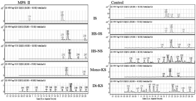
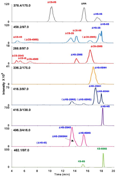
Glycosaminoglycans (GAGs) are long blocks of negatively charged polysaccharides. They are one of the major components of the extracellular matrix and play multiple roles in different tissues and organs. The accumulation of undegraded GAGs causes mucopolysaccharidoses (MPS). GAGs are associated with other pathological conditions such as osteoarthritis, inflammation, diabetes mellitus, spinal cord injury, and cancer. The need for further understanding of GAG functions and mechanisms of action boosted the development of qualitative and quantitative (alcian blue, toluidine blue, paper and thin layer chromatography, gas chromatography, high pressure liquid chromatography, capillary electrophoresis, 1,9-dimethylmethylene blue, enzyme linked-immunosorbent assay, mass spectrometry) techniques. The availability of quantitative techniques has facilitated translational research on GAGs into the medical field for: 1) diagnosis, monitoring, and screening for MPS; 2) analysis of GAG synthetic and degradation pathways; and 3) determination of physiological and pathological roles of GAGs. This review provides a history of development of GAG assays and insights about the use of tandem mass spectrometry and its applications for GAG analysis.

Keywords: Alcian blue; Chromatography, mucopolysaccharidoses; ELISA; Glycosaminoglycans; Mass spectrometry.


Conflict of interest statement

Conflict of interest: All the authors contributed to the Review Article and had no conflict of interest with any other party. Francyne Kubaski, Harumi Osago, Robert W. Mason, Seiji Yamaguchi, Hironori Kobayashi, Mikako Tsuchiya, Tadao Orii, and Shunji Tomatsu declare that they have no conflict of interests.

Figures

Fig. 1
Fig. 1
Structure of glycosaminoglycans. CS: chondroitin sulfate, DS: dermatan sulfate, HA: hyaluronic acid, HS: heparan sulfate, KS: keratan sulfate, UA: uronic acid, GlcA: glucuronic acid, Δ: unsaturated. Reproduced with permission from [ref. 152].
Fig. 2
Fig. 2
TB staining in growth plate of wild-type (left) and MPS VII (right) mice (12 weeks old). Chondrocytes in a wild-type mouse are stained while chondrocytes in MPS VII is ballooned and vacuolated.
Fig. 3
Fig. 3
AB staining for trachea with a 23-year-old MPS IVA patient and AB staining for electrophoretic urinary GAG from MPS IVA patients. Chondrocytes and their extracellular matrix as well as tracheal glands were stained with deep blue (left). (For interpretation of the references to color in this figure legend, the reader is referred to the web version of this article.)
Fig. 4
Fig. 4
Electrophoresis on a mono-dimensional run (left) and densitography (right) of urinary GAGs from MPS patients. Urinary GAGs was extracted by cetylpyridinium chloride (VPC) method and were separated by the electrophoresis. A mono-dimensional electrophoresis of urinary specimens from MPS I, II, III, IVA, VI, and VII patients and healthy control shows clear separation of specific GAGs (DS1, HS, DS2, C4S, C6S, and KS) (left). A healthy control sample yields C4S, C6S, and HS. Samples from MPS I and II patients provide more DS and HS. Samples from MPS III patients show a strong band of HS and there is no difference of the HS band between subclasses of MPS III. An MPS IVA sample provides a characteristic KS band while an MPS VI sample yields a thick DS band. Each separate GAG band is semi-quantified by Densitometer (right), and each type of MPS provides a unique pattern of densitography apart from the normal control pattern.
Fig. 5
Fig. 5
Multiple reaction monitoring (MRM) of DBS samples (control × MPS II patient). Chromatograms for disaccharides of chondrosine (IS), heparan sulfate (HS), mono-sulfated KS, di-sulfated KS. Equipment: 6460 Triple Quad MS/MS with 1260 infinity LC (Agilent Technologies). DBS: dried blood spot; IS: internal standard.
Fig. 6
Fig. 6
The extracted ion chromatogram of 23 disaccharides derived from four classes of GAGs by the LC/MS/MS analysis. The selected reaction monitoring transitions are shown in each chromatogram. The disaccharides shown in parentheses indicate the signals of their de-sulfated products by in-source fragmentation. Reproduced with permission from [ref. 152].

References

    1. Liu Z, Zhang F, Li L, Li G, He W, Lindhardt RJ. Compositional analysis and structural elucidation of glycosaminoglycans in chicken eggs. Glycoconj J. 2014;31:593–602. - PMC - PubMed
    1. Habuchi O. Diversity and functions of glycosaminoglycan sulfotransferases. Biochim Biophys Acta. 2000;1474:115–117. - PubMed
    1. Li G, Li L, Tian F, Zhang L, Xue C, Lindhardt RJ. Glycosaminoglycans of cultured cells using a rapid and sensitive LC–MS/MS approach. ACS Chem Biol. 2015;10:1303–1310. - PubMed
    1. Kiselova N, Dieker T, Spillmann D, Ramstrom M. An automated mass spectrometry-based screening method for analysis of sulfated glycosaminoglycans. Biochem Biophys Res Commun. 2014;450:598–603. - PubMed
    1. Kusche-Gullberg M, Khellén L. Sulfotransferases in glycosaminoglycanbiosynthesis. Curr Opin Struct Biol. 2003;13:605–611. - PubMed

MeSH terms

Substances