Comment
. 2016 Nov 14;30(5):683-693.
doi: 10.1016/j.ccell.2016.09.008. Epub 2016 Oct 13.

An LXR-Cholesterol Axis Creates a Metabolic Co-Dependency for Brain Cancers

Affiliations
Comment

An LXR-Cholesterol Axis Creates a Metabolic Co-Dependency for Brain Cancers

Genaro R Villa et al. Cancer Cell. .

Abstract

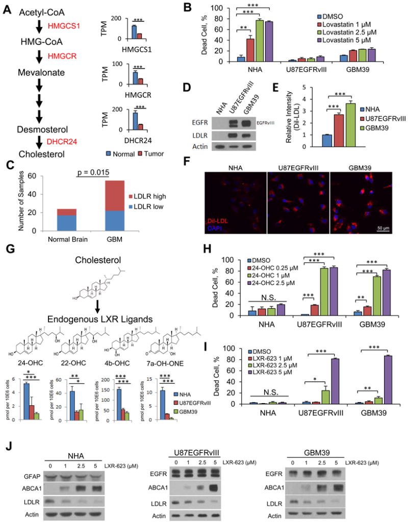
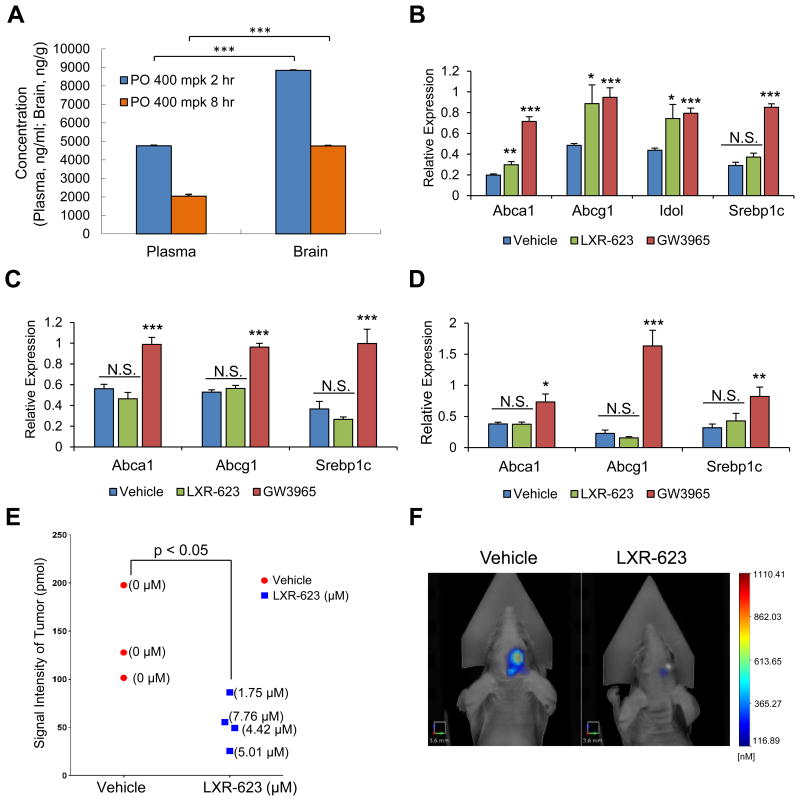
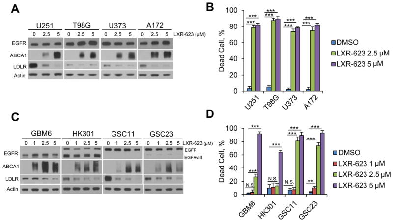
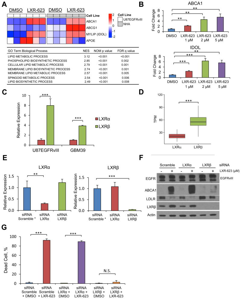
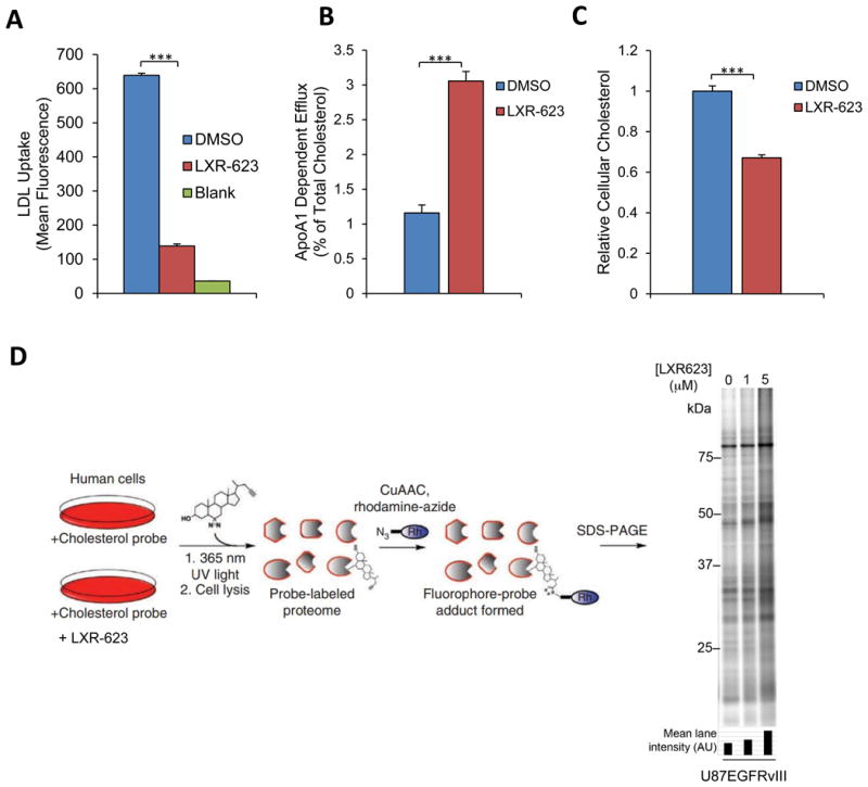
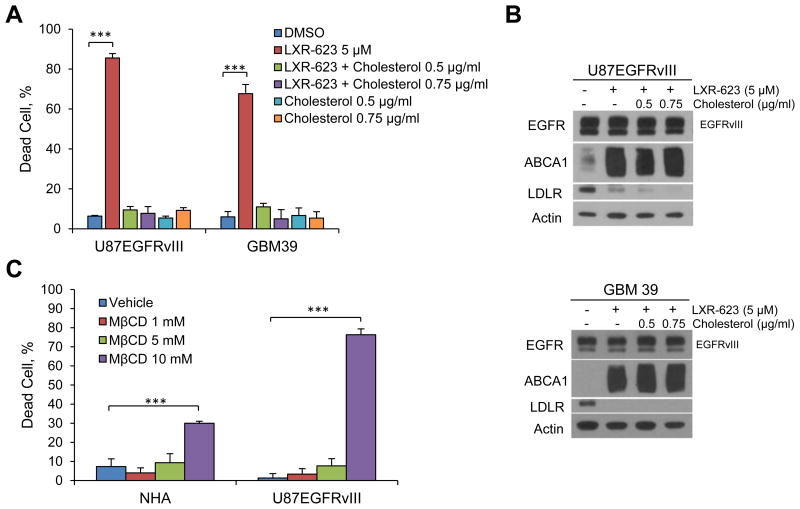
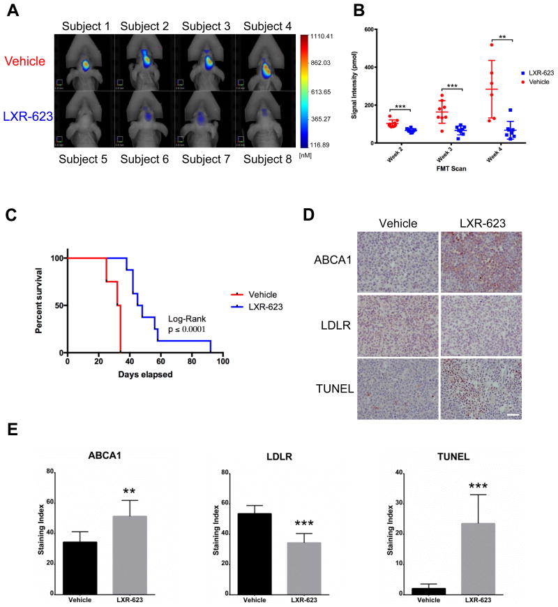
Small-molecule inhibitors targeting growth factor receptors have failed to show efficacy for brain cancers, potentially due to their inability to achieve sufficient drug levels in the CNS. Targeting non-oncogene tumor co-dependencies provides an alternative approach, particularly if drugs with high brain penetration can be identified. Here we demonstrate that the highly lethal brain cancer glioblastoma (GBM) is remarkably dependent on cholesterol for survival, rendering these tumors sensitive to Liver X receptor (LXR) agonist-dependent cell death. We show that LXR-623, a clinically viable, highly brain-penetrant LXRα-partial/LXRβ-full agonist selectively kills GBM cells in an LXRβ- and cholesterol-dependent fashion, causing tumor regression and prolonged survival in mouse models. Thus, a metabolic co-dependency provides a pharmacological means to kill growth factor-activated cancers in the CNS.

Keywords: brain cancer; cholesterol; glioblastoma; liver X receptor; metabolism; oxysterols.


Figures

Figure 1
Figure 1. Dysregulated cholesterol metabolism renders GBM cells selectively vulnerable to an exogenous LXR agonist
(A) Analysis of cholesterol synthesis genes in GBMs vs. normal brain from TCGA gene expression data. Data are reported as mean ± SEM. (B) FACS analysis of Annexin V/PI staining comparing NHA to U87EGFRvIII and GBM39 cells after a three-day treatment with the brain penetrant HMGCR inhibitor lovastatin. (C) Quantification of IHC analysis of tissue microarray samples for LDLR receptor. (D) Immunoblotting comparing EGFR and LDLR protein levels in NHA, U87EGFRvIII, and GBM39 cells. (E-F) FACS quantification (E) and representative images (F) of LDL uptake in U87EGFRvIII and GBM39 cells. (G) Schematic model and molecular structures of cholesterol synthesis into LXR ligands and LC/MS-MS data evaluating the levels of endogenous LXR ligands in GBM cells and astrocytes. Data are reported as mean ± SEM. (H) Quantification of cell death via FACS analysis of Annexin V/PI staining in response to endogenous LXR agonists at day five of treatment in NHA, U87EGFRvIII, and GBM39 cells. (I) Quantification of cell death in response to LXR-623 at day five of treatment in NHA, U87EGFRvIII, GBM39 cells. (J) NHA, U87EGFRvIII, and GBM39 were treated with the indicated concentrations of LXR-623 for 48 hr and immunoblotting was performed. Unless otherwise stated, data are reported as mean ± SD. *p < 0.05; **p < 0.01; ***p < 0.001; N.S. = not significant. See also Figure S1.
Figure 2
Figure 2. LXR-623 crosses the blood brain barrier, induces target gene expression, and achieves therapeutic levels in GBM cells in the brain with minimal activity in the periphery
(A) Mice were treated with a single dose of LXR-623 by oral gavage (PO) 400 mg/kg (mpk). Plasma and brain were extracted from mice at 2 or 8 hr after gavage. LXR-623 plasma concentrations are reported (n = 5 for each time point). (B-D) Mice were treated with LXR-623 or GW3965 (40 mg/kg) by oral gavage daily for three days. RNA was extracted from cerebral cortex (B), liver (C), and epididymal white adipose tissue, eWAT (D), and qPCR was performed for the indicated genes. n = 4 for each treatment condition. (E) U87EGFRvIII turbo FP 635 orthotopic mouse xenograft. Mice were treated with vehicle or LXR-623 400 mpk PO daily. Tumor size was assessed via fluorescence molecular tomography (FMT) on day five of treatment. Tumors were excised from mice and the intratumor concentration of LXR-623 was assessed via liquid chromatography-tandem mass spectrometry (LC-MS/MS). Intratumoral LXR-623 concentrations are indicated within parentheses. n = 3 for vehicle and n = 4 for LXR-623 treated mice. (F) Representative FMT images of mice from (E). Scale bar, 3.6 mm. Data are reported as mean ± SEM. *p < 0.05; **p < 0.01; ***p < 0.001; N.S. = not significant. See also Figure S2.
Figure 3
Figure 3. LXR-623 induces cell death in established and patient-derived GBM cells
(A) Established GBM cell lines U251, T98G, U373 and A172 were treated with the indicated concentration of LXR-623 for 48 hr and immunoblotting was performed with the indicated antibodies. (B) Trypan blue exclusion assay was carried out in parallel to (A) after three days of LXR-623 treatment. (C) Patient derived GBM neurosphere lines GBM6, HK301, GSC11 and GSC23 were treated with the indicated concentration of LXR-623 for 48 hr and immunoblotting was performed as in (A). (D) Trypan blue exclusion assay of cells from (C) after five days of treatment. Data are reported as mean ± SD. **p < 0.01; ***p < 0.001; N.S. = not significant. See also Figure S3.
Figure 4
Figure 4. LXR-623 kills GBM cells through activation of LXRβ, the dominant subtype in brain tumors
(A) Microarray analysis of LXR target genes in U87EGFRvIII and NHA cells treated with LXR-623 5 μM for 24 hr. Triplicates are shown. Gene Set Enrichment Analysis (GSEA) for Gene Ontology (GO) pathways for microarray data in (A) shown in the table below. (B) U87EGFRvIII cells were treated with LXR-623 for 48 hr and qPCR was performed for LXR target genes. (C) RNA was extracted from U87EGFRvIII, GBM39, and qPCR was performed for LXRα and LXRβ. (D) Boxplot of The Cancer Genome Atlas (TCGA) RNASeq data from patients with GBM showing LXRβ is the primary LXR subtype expressed in GBM. The box extends from the 25th to 75th percentiles and the middle line inside indicates median. Whiskers are drawn down to the 10th percentile and up to the 90th. (E) U87EGFRvIII cells were transfected with siRNA pools targeting LXRα or LXRβ. After 48 hr RNA was extracted and qPCR was performed. *Gene expression was normalized for scramble control. (F) Cells were transfected with siRNA as in (E) and treated with LXR-623 for 24 hr. Immunoblotting was performed with the indicated antibodies. (G) U87EGFRvIII cells were transfected with siRNA as in (E) and treated with LXR-623. Trypan blue exclusion assay was performed after three days of treatment. Data are reported as mean ± SD. **p < 0.01; ***p < 0.001; N.S. = not significant. See also Figure S4.
Figure 5
Figure 5. LXR-623 Depletes GBM Cells of cholesterol
(A) U87EGFRvIII cells were treated with LXR-623 for a total of 48 hr. Cells were incubated with fluorescently labeled LDL for four hr and LDL uptake was determined via flow cytometry. (B) U87EGFRvIII cells were loaded with 3H-cholesterol and treated with LXR-623 5 μM. Cholesterol efflux was determined by scintillation counting. (C) U87EGFRvIII cells were treated as in (A) and total cholesterol levels were assessed by LC/MS. (D) Sterol probe labeling profile (10 μM probe, 30 min) of U87EGFRvIII cells pre-treated with DMSO or the indicated concentrations of LXR-623 for 48 hr. Data are reported as mean ± SD. ***p < 0.001. See also Figure S5.
Figure 6
Figure 6. LXR-623 kills GBM Cells by Depleting Cholesterol
(A) U87EGFRvIII and GBM39 cells were treated with LXR-623 in the presence or absence of methyl-β-cyclodextrin complexed to cholesterol at the indicated concentrations (final concentration of cholesterol is shown). Cell death was assessed via Annexin/PI staining on day three for U87EGFRvIII and via trypan blue staining on day five for GBM39. (B) U87EGFRvIII (top panel) or GBM39 (bottom panel) were treated with LXR-623 for 48 hr in the presence of absence of MβCD-Cholesterol. Immunoblotting was performed with the indicated antibodies. (C) NHA and U87EGFRvIII were treated with the indicated concentrations of methyl-β-cyclodextrin (MβCD) for 1 hr and cell death was assessed by trypan blue exclusion assay. Data are reported as mean ± SD. ***p < 0.001. See also Figure S6.
Figure 7
Figure 7. LXR-623 inhibits tumor growth, promotes tumor cell death and prolongs the survival of mice bearing intracranial patient-derived GBMs
(A) GBM39 patient derived neurosphere cells, engineered to stably express the infrared protein 720 (IRFP 720), were orthotopically injected into five-week old nu/nu mice. Mice were treated with vehicle or LXR-623 400 mpk PO daily (n = 8 for each group). Representative FMT images of mice at week five of treatment. Scale bar, 3.8 mm. (B) Tumor size was assessed via fluorescence molecular tomography (FMT) weekly. (C) Kaplan-Meier curves assessing overall survival of mice from (A). Log-rank (Mantel-Cox) test: p = 0.0001, Gehan-Breslow-Wilcoxon test: p = 0.0002. (D) Tumors were excised and immunohistochemistry analysis was performed with the indicated antibodies. Scale bar, 50 μm. (E) Quantification of the immunohistochemistry performed in (D). Data are reported as mean ± SEM. **p < 0.01; ***p < 0.001. See also Figure S7.

Comment on

References

    1. Björkhem I. Crossing the barrier: oxysterols as cholesterol transporters and metabolic modulators in the brain. J Intern Med. 2006;260:493–508. - PubMed
    1. Björkhem I, Meaney S. Brain cholesterol: long secret life behind a barrier. Arterioscler Thromb Vasc Biol. 2004;24:806–815. - PubMed
    1. Bovenga F, Sabbà C, Moschetta A. Uncoupling nuclear receptor LXR and cholesterol metabolism in cancer. Cell Metab. 2015;21:517–526. - PubMed
    1. Bradley MN, Hong C, Chen M, Joseph SB, Wilpitz DC, Wang X, Lusis AJ, Collins A, Hseuh WA, Collins JL, et al. Ligand activation of LXR beta reverses atherosclerosis and cellular cholesterol overload in mice lacking LXR alpha and apoE. J Clin Invest. 2007;117:2337–2346. - PMC - PubMed
    1. Brastianos PK, Carter SL, Santagata S, Cahill DP, Taylor-Weiner A, Jones RT, Van Allen EM, Lawrence MS, Horowitz PM, Cibulskis K, et al. Genomic Characterization of Brain Metastases Reveals Branched Evolution and Potential Therapeutic Targets. Cancer Discov. 2015;5:1164–1177. - PMC - PubMed

Substances