. 2016 Dec;186(12):3066-3073.
doi: 10.1016/j.ajpath.2016.07.026. Epub 2016 Oct 13.

Lymphoid Aggregates Remodel Lymphatic Collecting Vessels that Serve Mesenteric Lymph Nodes in Crohn Disease

Affiliations

Lymphoid Aggregates Remodel Lymphatic Collecting Vessels that Serve Mesenteric Lymph Nodes in Crohn Disease

Gwendalyn J Randolph et al. Am J Pathol. 2016 Dec.

Abstract

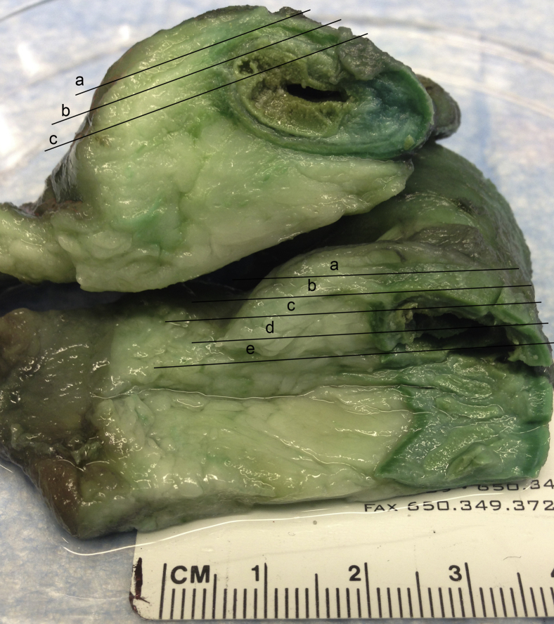
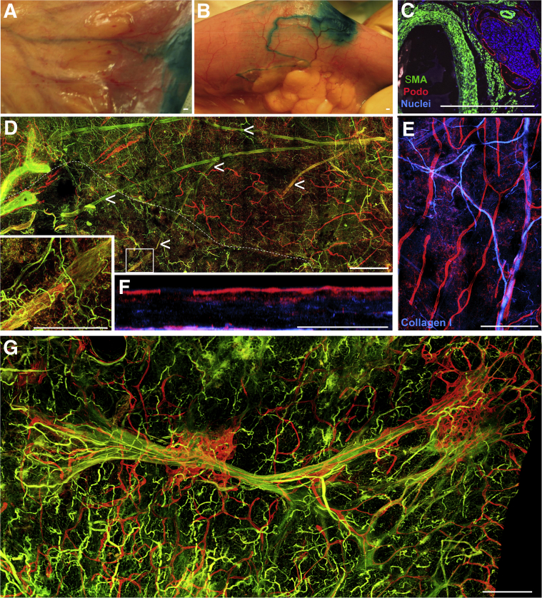
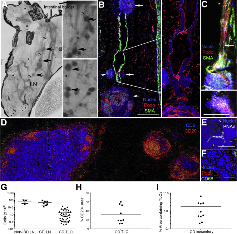
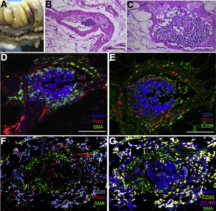
Early pathological descriptions of Crohn disease (CD) argued for a potential defect in lymph transport; however, this concept has not been thoroughly investigated. In mice, poor healing in response to infection-induced tissue damage can cause hyperpermeable lymphatic collecting vessels in mesenteric adipose tissue that impair antigen and immune cell access to mesenteric lymph nodes (LNs), which normally sustain appropriate immunity. To investigate whether analogous changes might occur in human intestinal disease, we established a three-dimensional imaging approach to characterize the lymphatic vasculature in mesenteric tissue from controls or patients with CD. In CD specimens, B-cell-rich aggregates resembling tertiary lymphoid organs (TLOs) impinged on lymphatic collecting vessels that enter and exit LNs. In areas of creeping fat, which characterizes inflammation-affected areas of the bowel in CD, we observed B cells and apparent innate lymphoid cells that had invaded the lymphatic vessel wall, suggesting these cells may be mediators of lymphatic remodeling. Although TLOs have been described in many chronic inflammatory states, their anatomical relationship to preestablished LNs has never been revealed. Our data indicate that, at least in the CD-affected mesentery, TLOs are positioned along collecting lymphatic vessels in a manner expected to affect delivery of lymph to LNs.


Figures

Figure 1
Figure 1
Depiction of tissue slicing performed to generate material for whole mount imaging. Two segments of ileum are stacked on each other with intestinal lumen on the right and mesenteric fat on the left. The black lines indicate the typical locations at which slices, each labeled a lower case letter ae, were made for analysis. Slices labeled a were those that contained serosal epithelium and thus contained subserosal lymphatic capillaries. Most collecting vessels were found in slices a or b.
Figure 2
Figure 2
Characterization of the human mesenteric lymphatic vasculature in healthy and Crohn disease (CD)-affected tissue. A and B: Representative images of small bowel loops after injection of patent blue dye in the submucosa during surgical resection. A: An area that showed no evidence of disease. B: An area affected by the development of creeping fat during the course of progressive CD. C: Immunofluorescence analysis that depicts collections of lymphatic vessels in the human CD-affected mesentery nearby blood vessels, with patient referred to surgery because of ileal stenosis from stricturing disease. The lymphatic vessels [podoplanin (Podo), red] encase a collection of nucleated cells stained with DAPI (blue). α-Smooth muscle actin (α-SMA; green) staining failed to identify collecting lymphatic vessels (podoplanin-positive endothelium with α-SMA–positive cells surrounding them) in most paraffin-embedded cross sections. D: Whole mount confocal imaging of large area of mesenteric adipose tissue (ie, cm × cm × mm scale) from a control specimen unaffected by inflammatory bowel disease (IBD) reveals blinded-ended lymphatic capillaries (red) and other lymphatic capillaries near α-SMA–positive blood vessels (left along main image). Tissue depicted below the dotted white line represents an area with mesothelial layer removed. Inset: Region of enlargement, in which the presence of a collecting lymphatic vessel (red + green) is evident. Arrowheads trace this collecting lymphatic vessel branching structure throughout the tissue. E and F: Collagen I (blue) and podoplanin (red) staining depict blood and lymphatic capillaries, respectively, from a control (no IBD) sample distinct from that shown in D. F: A region from E turned to the side to reveal major lymphatic capillary network (red) just below the mesothelial surface and a second lymphatic capillary grouping around the deeper blood vessel. G: Confocal scan of a large area (ie, cm × cm × mm) from a CD-affected mesentery stained for podoplanin (red) and α-SMA (green). Lymphatic capillaries are markedly expanded around the larger blood vessel. The right edge of the image marks the border of the image that was nearest the intestinal wall.
Figure 3
Figure 3
Improved method to identify tissue specimens containing lymphatic collecting vessels links them to accumulation of tertiary lymphoid organs (TLOs) in Crohn disease (CD) mesentery. In all parts with the exception of D, the images are positioned so that the intestinal border is toward the top of the figure. In D, the intestinal border is on the right of the image. A: Evaluation of methyl salicylate–cleared tissue slices using a light-emitting diode (LED) allows detection of lymph nodes (LNs), lymphoid follicles, and vessels that link them together. Three CD mesenteric specimens shown herein arise from patients with ileal disease that led to surgery because of ileal occlusion in one case, stenosis in another, and fistulizing, perforating disease in the third. Image on the left shows LN with collecting lymphatic vessel (arrowheads) leading back to intestinal wall, with smaller lymphoid collections along the route (arrows). Two images on the right show chains of TLOs that are too small to be visible in the tissue slice when examined by eye, but which are readily identified using LED light scanning. B: Whole mount samples as in A were processed to generate 5-μm paraffin-embedded sections that were immunostained to identify α-smooth muscle actin (α-SMA)–positive vessels and podoplanin (Podo). Data reveal collecting lymphatic vessels interrupted by lymphoid follicle structures (white arrows). Enlargement on the right allows a better view, in the absence of the strong α-SMA staining, of the weaker podoplanin staining that characterizes the lumen of collecting lymphatic vessels. C: Different patient mesenteric fat sample cut in thicker 100-μm sections showing a collecting lymphatic vessel running into a follicle, with some tissue damage generated during delicate preparation. White arrow points to lymphatic collecting vessel valve that defines the direction of flow as being toward the follicle. The lower image, the next 100-μm section in the sequence, shows lymphatic vessels as they wind their way through to the efferent side of the follicle. D: CD20 and CD3 staining to identify follicles as lymphocyte-rich structures. LN on the left, two follicles on the right. Collecting lymphatic vessel that links these structures in a chain is thin enough that it is missing in this 5-μm paraffin section. E and F: Staining of follicle structures for peripheral node addressin (PNAd; E) and CD68 (F). Asterisks in E denote adipocytes. G: Quantification of cell numbers in lymph nodes and TLOs. Each lymph node analysis comes from a different CD specimen. For the TLO analysis, we measured the size of multiple TLOs in the same specimen. The 41 TLO evaluations depicted in the panel arise from TLOs combined from 10 different patients. H: Tissues from nine of the patient samples were stained for CD20, and the fraction of TLO structures positive for CD20 is depicted. I: The TLO area in the mesentery of the same 10 CD patients was quantified relative to the total mesenteric area in cross sections. Each dot in H and I represents data from one patient. Horizontal lines represent the mean value in the respective data column. Scale bars = 300 μm.
Figure 4
Figure 4
B cells and IL-33R+ CD3 cells localize to the collecting lymphatic vessel walls associated with creeping fat. A: Portion of the small bowel bearing relocalized, or creeping, fat from a patient receiving ileal resection because of Crohn disease complications. Adipose tissue was cut so as to bisect lobes of the fat, in the opposite orientation to the usual direction we cut the tissue (Figure 1). B and C: Hematoxylin and eosin sections from these lobes showed the presence of collecting lymphatic vessels. The two parts show the same vessel at different points. D and E: Adjacent sections from the block used in C were stained to identify nucleated cells, podoplanin (Podo), and α-smooth muscle actin (α-SMA; D) or CD3, CD20, and IL-33R (E). F and G: Analysis of a tertiary lymphoid organ from different patient as above. Seven-color confocal scanning was performed with the display of data in two parts. Scale bars = 50 μm.

References

    1. de Souza H.S., Fiocchi C. Immunopathogenesis of IBD: current state of the art. Nat Rev Gastroenterol Hepatol. 2016;13:13–27. - PubMed
    1. Fonseca D.M., Hand T.W., Han S.J., Gerner M.Y., Glatman Zaretsky A., Byrd A.L., Harrison O.J., Ortiz A.M., Quinones M., Trinchieri G., Brenchley J.M., Brodsky I.E., Germain R.N., Randolph G.J., Belkaid Y. Microbiota-dependent sequelae of acute infection compromise tissue-specific immunity. Cell. 2015;163:354–366. - PMC - PubMed
    1. Mikulski Z., Johnson R., Shaked I., Kim G., Nowyhed H., Goodman W., Chodaczek G., Pizarro T.T., Cominelli F., Ley K. SAMP1/YitFc mice develop ileitis via loss of CCL21 and defects in dendritic cell migration. Gastroenterology. 2015;148:783–793e5. - PMC - PubMed
    1. Van Kruiningen H.J., Colombel J.F. The forgotten role of lymphangitis in Crohn's disease. Gut. 2008;57:1–4. - PubMed
    1. von der Weid P.Y., Rehal S., Ferraz J.G. Role of the lymphatic system in the pathogenesis of Crohn's disease. Curr Opin Gastroenterol. 2011;27:335–341. - PubMed

Publication types

MeSH terms