. 2015 Feb;103(2):205-224.
doi: 10.1109/JPROC.2014.2385105. Epub 2015 Mar 24.

Biomedical Applications of Untethered Mobile Milli/Microrobots

Affiliations

Biomedical Applications of Untethered Mobile Milli/Microrobots

Metin Sitti et al. Proc IEEE Inst Electr Electron Eng. 2015 Feb.

Abstract

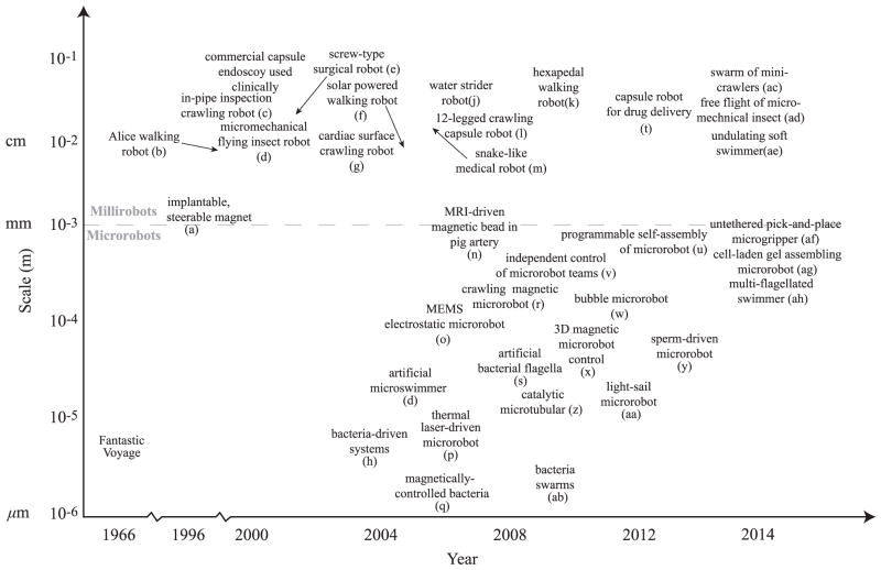
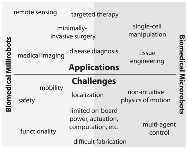
Untethered robots miniaturized to the length scale of millimeter and below attract growing attention for the prospect of transforming many aspects of health care and bioengineering. As the robot size goes down to the order of a single cell, previously inaccessible body sites would become available for high-resolution in situ and in vivo manipulations. This unprecedented direct access would enable an extensive range of minimally invasive medical operations. Here, we provide a comprehensive review of the current advances in biome dical untethered mobile milli/microrobots. We put a special emphasis on the potential impacts of biomedical microrobots in the near future. Finally, we discuss the existing challenges and emerging concepts associated with designing such a miniaturized robot for operation inside a biological environment for biomedical applications.

Keywords: Biomedical engineering; medical robots; microrobots; minimally invasive surgery.


Figures

Fig. 1
Fig. 1
Approximate timeline showing the emerging new milli/microrobot systems with their given overall size scale as significant milestones. (a) Implantable tiny permanent magnet steered by external electromagnetic coils [16]. (b) Alice 1 cm3 walking robot [17]. (c) In-pipe inspection crawling robot [18]. (d) Micromechanical flying insect robot [19]. (e) Screw-type surgical millirobot [20]. (f) Solar powered walking robot [21]. (g) Cardiac surface crawling medical robot [22]. (h) Bacteria-driven biohybrid microrobots [23]. (i) biohybrid magnetic microswimmer [24]. (j) Water strider robot [25]. (k) Hexapedal compliant walking robot [26]. (l) 12-legged crawling capsule robot [27]. (m) Snake-like medical robot [28]. (n) Magnetic bead driven by a Magnetic Resonance Imaging device in pig artery [29]. (o) MEMS electrostatic microrobot [30]. (p) Thermal laser-driven microrobot [31]. (q) Magnetically controlled bacteria [32]. (r) Crawling magnetic microrobot [33]. (s) Magnetic microswimmer inspired by bacterial flagella [34], [35]. (t) Flexible capsule endoscope with drug delivery mechanism [36]. (u) Programmable self-assembly of microrobots [37]. (v) Independent control of microrobot teams [38]. (w) Bubble microrobot [39]. (x) 3D magnetic microrobot control [40]. (y) Sperm-driven biohybrid microrobot [41]. (z) Catalytic microtubular [42]. (aa) Light-sail microrobot [43]. (ab) Bacteria swarms as microrobotic manipulation systems [44]. (ac) Swarm of mini-crawlers [45]. (ad) Free flight of micromechanical insect [46]. (ae) Undulating soft swimmer [47]. (af) Untethered pick-and-place microgripper [48]. (ag) Cell-laden gel assembling microrobot [49]. (ah) Multiflagellated swimmer [50].
Fig. 2
Fig. 2
Some existing off-board approaches to mobile microrobot actuation and control in 2D. (a) Magnetically driven crawling robots include the Mag-μBot [33], the Mag-Mite magnetic crawling microrobot [59], the magnetic microtransporter [60], rolling magnetic microrobot [61], the diamagnetically-levitating mm-scale robot [62], the self-assembled surface swimmer [63], and the magnetic thin-film microrobot [64]. (b) Thermally driven microrobots include the laser-activated crawling microrobot [31], microlight sailboat [43], and the optically controlled bubble microrobot [39]. (c) Electrically driven microrobots include the electrostatic scratch-drive microrobot [65] and the electrostatic microbiorobot [60]. Other microrobots which operate in 2D include the piezoelectric-magnetic microrobot MagPieR [66] and the electrowetting droplet microrobot [67].
Fig. 3
Fig. 3
Some existing off-board and on-board approaches to mobile milli/microrobot actuation and control in 3D. (a) Chemically propelled designs include the microtubular jet microrobot [42] and the electro-osmotic swimmer [68]. (b) Swimming milli/microrobots include the colloidal magnetic swimmer [24], the magnetic thin-film helical swimmer [69], the micron-scale magnetic helix fabricated by glancing angle deposition [35], the microhelix microrobot with cargo carrying cage, fabricated by direct laser writing [70] and the microhelix microrobot with magnetic head, fabricated as thin-film and rolled using residual stress [34]. (c) Milli/microrobots pulled in 3D using magnetic field gradients include the nickel microrobot capable of five-degrees-of-freedom (DOF) motion in 3D using the OctoMag system [40] and the MRI-powered and imaged magnetic bead [71]. (d) Cell-actuated biohybrid approaches include the artificially-magnetotactic bacteria [72], the cardiomyocyte driven microswimmers [73], the chemotactic steering of bacteria-propelled microbeads [74], sperm-driven and magnetically steered microrobots [41], and the magnetotactic bacteria swarm manipulating microscale bricks [44].
Fig. 4
Fig. 4
Applications and challenges for biomedical milli/microrobots.
Fig. 5
Fig. 5
(a) Photograph of the prototype (left picture) of an example magnetically actuated capsule millirobot for active imaging inside stomach. A CMOS camera and LED lighting were integrated to the soft capsule robot, which can axially deform due to external magnetic actuation control. (b) An active imaging example (right picture) snapshot of the surgical stomach model from the CMOS camera during its active orientation control by an external magnet.
Fig. 6
Fig. 6
Active drug delivery demonstration of a soft capsule millirobot (see Table 1 for its specifications) inside stomach. (a)–(c) Time snapshots of the drug diffusion during the active compression of the drug chamber with the remote magnetic actuation [92].
Fig. 7
Fig. 7
Conceptual sketch of a bacteria-propelled biohybrid microrobot swarm, as a dense stochastic network, transporting and delivering drugs on targeted regions inside the stagnant fluid regions of the human body.
Fig. 8
Fig. 8
3D assembly of cell-laden microgels by different microrobots. (a) Magnetic crawling microrobot [49]. The microgel is pushed by the microrobot to the desired position. A microfabricated ramp is used to elevate the microrobot to higher layer of the tissue constructs. (b) Magnetic microgripper [48]. The jaw is opened and closed by external magnetic field to pick up and release the microobject. (c) Magnetic microrobot with bubble capillary gripper [117]. Changing the pressure inside the working environment can extend and retract the bubble to pick up and release the microobject. (d) Magnetic coil system for microrobot control.
Fig. 9
Fig. 9
(a)–(f) Capillary gripping microrobot manipulating hydrogels into a stack as shown from the top-down view. (a) The microrobot position is given by the red cross and the desired position is given by the blue cross. microrobot position control is achieved by a PID controller used to determine the applied magnetic force. The hydrogels are the three circular disks (diameter ~ 350 μm) and the microrobot is a capillary gripping microrobot with a side dimension of 150 μm. (b) The microrobot is directed above the hydrogel and the bubble is drawn out of the cavity by a negative applied pressure in the microrobot workspace. The microrobot is then lowered onto the hydrogel. (c) The microrobot with the hydrogel positions itself over the center hydrogel and comes into contact. (d) The microrobot detaches from the stack of two hydrogels. (e), (f) The process is repeated for the left hydrogel, resulting in a three-hydrogel stack. Scale bar is 1 mm. (g) Example magnetic microrobot with a cavity for bubble-based capillary gripping. The four cones ensure surface contact is minimized when releasing parts. (h) Peel off force versus the average bubble height. The peel off force is calculated as the equivalent force acting on the center of the microrobot due to the applied magnetic torque. The magnetic torque is calculated from the applied uniform magnetic field and the known magnetization of the microrobot. The bubble height is measured from the cavity opening to the highest point of the bubble when it is not in contact with the test substrate. The height of 0 indicates the bubble is completely inside the cavity and there should be no capillary attachment force and is considered to be in the “release” state. Any positive non-zero bubble height will be considered the “pick” state. On a test silicon substrate, the best current work shows an attachment switching ratio of peel off force in the “pick” state to the peel off force in the “release” state of 25 : 1.
Fig. 10
Fig. 10
Conceptual figure/illustration showing all potential applications of microrobotic cell manipulation.
Fig. 11
Fig. 11
Visionary design of a soft, modular microrobot with spatio-selective functionalization. Each functional component is assembled on a main board. The main board further serves as a large depot for therapeutics to launch controlled release at the site of action. A closed-loop autonomous locomotion (e.g., a biohybrid design) couples environmental signals to motility. Targeting units enable reaching and localization at the intended body site. MRI contrast agents loaded on the microrobot enables visualization as well as manual steering on demand. Gold nanorods enable plasmonic heating to decompose a tumor tissue.
Fig. 12
Fig. 12
Conceptual sketch of a large number of microrobots made of smart materials that can be remotely actuated and controlled inside the human body with a user interface to achieve different biomedical functions.

References

    1. Sitti M. Miniature devices: Voyage of the micro-robots. Nature. 2009 Apr;458:1121–1122. - PubMed
    1. Nelson BJ, Kaliakatsos IK, Abbott JJ. Micro-robots for minimally invasive medicine. Annu Rev Biomed Eng. 2010 Aug;12:55–85. - PubMed
    1. Hale MF, Sidhu R, McAlindon ME. Capsule endoscopy: Current practice and future directions. World J Gastroenterol. 2014 Jun;20:7752–7759. - PMC - PubMed
    1. Liao Z, Gao R, Xu C, Li ZS. Indications and detection, completion, retention rates of small-bowel capsule endoscopy: A systematic review. Gastrointest Endosc. 2010 Feb;71:280–286. - PubMed
    1. Li Z, Liao Z, McAlindon M. Handbook of Capsule Endoscopy. Dordrecht, The Netherland: Springer Netherlands; 2014.

LinkOut - more resources