

Radiologic and histological observations in experimental T1-T12 dorsal arthrodesis: A qualitative description of T1-T12 segment and other body parts involved, between prepubertal age and skeletal maturityxs
- PMID: 27746501
- PMCID: PMC5017180
- DOI: 10.4103/0019-5413.189600
Radiologic and histological observations in experimental T1-T12 dorsal arthrodesis: A qualitative description of T1-T12 segment and other body parts involved, between prepubertal age and skeletal maturityxs
Abstract
Background: This experimental study provides a qualitative description and the morpho-structural features of the fusions taking place in the thoracic spine between prepubertal age and skeletal maturity. There is a lack of informations regarding the influence of partial or total dorso-thoracic vertebral arthrodesis on the development of the thoracic cage as well as its potential effects on different intra and extra-thoracic organs. This study admits the hypothesis that vertebral arthrodesis may have influence on other body areas and so, it intends to verify the possible secondary involvement of other body parts, such as intervertebral discs, cervical and thoracic spinal ganglia, sternocostal cartilage, ovaries and lungs.
Materials and methods: Fifty-four female New Zealand white rabbits were submitted to dorsal arthrodesis. The radiologic imaging and light microscopy histological pictures were taken and studied in all. Computed tomography (CT) scan measurements were performed in operated and sham operated rabbits at different time. Similarly, histological specimens of intervertebral discs, cervical and thoracic spinal ganglia, sternocostal cartilage, ovaries and lungs were analyzed at different times. The study ended at the age of 17-18 months.
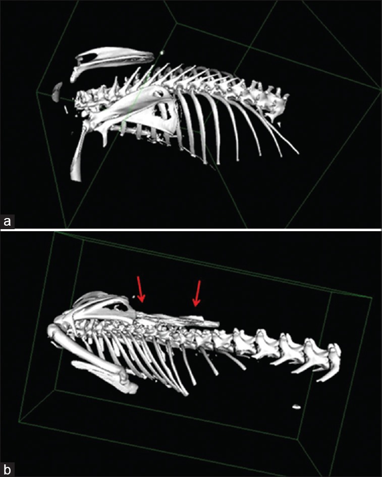
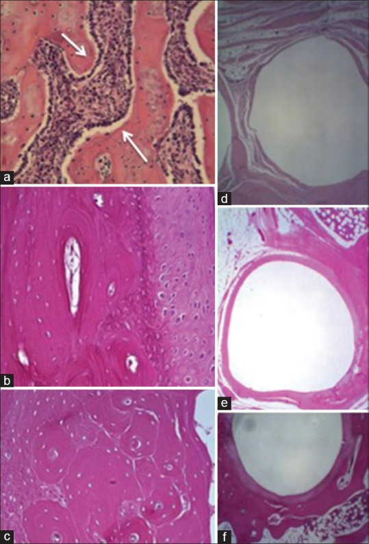
Results: Most rabbits had formed a fusion mass, which was only fibrous at first, then osteofibrous and finally, in the older subjects, structured in lamellar-osteon tissue. Intervertebral foramens were negatively involved in vertebral arthrodesis, as shown by CT scans. Intervertebral discs showed irregular aspects. The increase of atresic follicles and the reduction of primordial follicles in operated rabbits led to the hypothesis of a cause-effect relationship between arthrodesis and modified hormonal status. Dorsal root ganglia showed microscopic alterations in operated rabbits especially.
Conclusions: The process of fusion mass and bone formation, associated with the arthrodesis, involves at different degrees of the vertebral bodies, discs and intervertebral foramens, ganglia and spinal nerve roots.
Keywords: Computed tomography scan images; Spinal column; arthrodesis; computed tomography scanners; dorsal arthrodesis; histology; histopathology; prepubertal rabbits; qualitative study; rabbits.
Figures


References
-
- Hibbs R. A report of 59 cases of scoliosis treated by fusion operation. J Bone Joint Surg Am. 1924;6:3–37.
-
- Harrington PR. Treatment of scoliosis. Correction and internal fixation by spine instrumentation. J Bone Joint Surg Am. 1962;44-A:591–610. - PubMed
-
- Risser JC. Treatment of scoliosis during the past 50 years. Clin Orthop Relat Res. 1966;44:109–13. - PubMed
-
- Luque ER. The anatomic basis and development of segmental spinal instrumentation. Spine (Phila Pa 1976) 1982;7:256–9. - PubMed
-
- Cotrel Y, Dubouset J. A new posterior segmental vertebral arthrodesis technique. Rev Chir Orthop. 1984;70:489–99. - PubMed
LinkOut - more resources
Full Text Sources
Other Literature Sources