Review
. 2016 Sep 29:10:76.
doi: 10.3389/fncir.2016.00076. eCollection 2016.

Somatostatin-Expressing Inhibitory Interneurons in Cortical Circuits

Affiliations
Review

Somatostatin-Expressing Inhibitory Interneurons in Cortical Circuits

Iryna Yavorska et al. Front Neural Circuits. .

Abstract

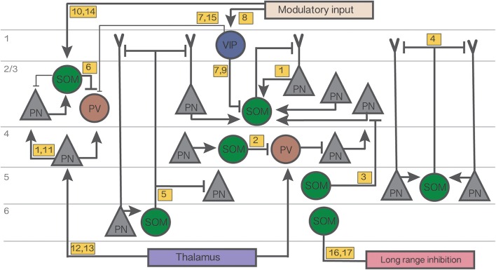
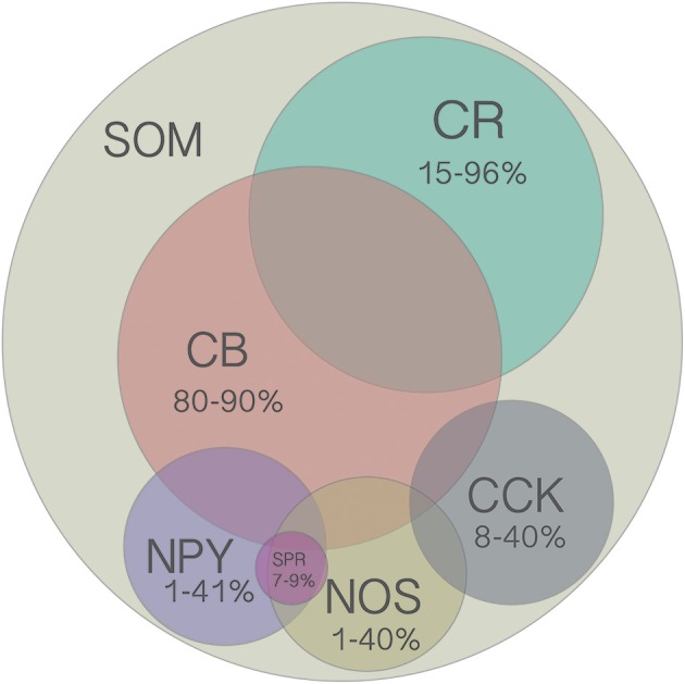
Cortical inhibitory neurons exhibit remarkable diversity in their morphology, connectivity, and synaptic properties. Here, we review the function of somatostatin-expressing (SOM) inhibitory interneurons, focusing largely on sensory cortex. SOM neurons also comprise a number of subpopulations that can be distinguished by their morphology, input and output connectivity, laminar location, firing properties, and expression of molecular markers. Several of these classes of SOM neurons show unique dynamics and characteristics, such as facilitating synapses, specific axonal projections, intralaminar input, and top-down modulation, which suggest possible computational roles. SOM cells can be differentially modulated by behavioral state depending on their class, sensory system, and behavioral paradigm. The functional effects of such modulation have been studied with optogenetic manipulation of SOM cells, which produces effects on learning and memory, task performance, and the integration of cortical activity. Different classes of SOM cells participate in distinct disinhibitory circuits with different inhibitory partners and in different cortical layers. Through these disinhibitory circuits, SOM cells help encode the behavioral relevance of sensory stimuli by regulating the activity of cortical neurons based on subcortical and intracortical modulatory input. Associative learning leads to long-term changes in the strength of connectivity of SOM cells with other neurons, often influencing the strength of inhibitory input they receive. Thus despite their heterogeneity and variability across cortical areas, current evidence shows that SOM neurons perform unique neural computations, forming not only distinct molecular but also functional subclasses of cortical inhibitory interneurons.

Keywords: GIN; Martinotti; VIP; disinhibitory; parvalbumin; somatostatin; x94; x98.


Figures

Figure 1
Figure 1
Summary diagram of the cortical circuits in which SOM cells participate. Numbers in yellow boxes refer to the references below. Some microcircuits combine results from different studies and include speculative connections. Connections not mentioned in the main text or not pertaining to SOM networks are omitted for simplicity. Thin lines indicate weaker connections. Figure references: 1. Adesnik et al. (2012); 2. Xu et al. (2013); 3. Kapfer et al. (2007); 4. Silberberg and Markram (2007); 5. Wang et al. (2004); 6. Cottam et al. (2013); 7. Pi et al. (2013); 8. Fu et al. (2014); 9. Karnani et al. (2016a); 10. Kawaguchi et al. (1997); 11. Li et al. (2015); 12. Cruikshank et al. (2010); 13. Beierlein et al. (2003); 14. Fanselow et al. (2008); 15. Letzkus et al. (2011); 16. Tomioka et al. (2005); 17. Endo et al. (2016).
Figure 2
Figure 2
Venn diagram of all molecular markers that are colocalized with SOM (CB, CR, NPY, NOS, CCK). Numbers indicate the range of reported percentages of SOM-positive cells that coexpress a given marker across studies in different species (mouse or rat), cortical layer (2–6), and cortical region (frontal, visual, or somatosensory). For more details, see Table 2. Area of each circle approximately represents the average range. Overlap of circles indicates known coexpression of depicted markers, however the area of the overlap does not indicate the extent of coexpression. Although there are no reports of SOM cells coexpressing more than two other molecular markers, this possibility has not been ruled out since it was rarely tested for.

Similar articles

Cited by

References

    1. Adesnik H., Bruns W., Taniguchi H., Huang Z. J., Scanziani M. (2012). A neural circuit for spatial summation in visual cortex. Nature 490, 226–231. 10.1038/nature11526 - DOI - PMC - PubMed
    1. Amitai Y., Gibson J. R., Beierlein M., Patrick S. L., Ho A. M., Connors B. W., et al. . (2002). The spatial dimensions of electrically coupled networks of interneurons in the neocortex. J. Neurosci. 22, 4142–4152. - PMC - PubMed
    1. Anderson J. S., Carandini M., Ferster D. (2000). Orientation tuning of input conductance, excitation, and inhibition in cat primary visual cortex. J. Neurophysiol. 84, 909–926. - PubMed
    1. Ascoli G. A., Alonso-Nanclares L., Anderson S. A., Barrionuevo G., Benavides-Piccione R., Burkhalter A., et al. . (2008). Petilla terminology: nomenclature of features of GABAergic interneurons of the cerebral cortex. Nat. Rev. Neurosci. 9, 557–568. 10.1038/nrn2402 - DOI - PMC - PubMed
    1. Atallah B. V., Bruns W., Carandini M., Scanziani M. (2012). Parvalbumin-expressing interneurons linearly transform cortical responses to visual stimuli. Neuron 73, 159–170. 10.1016/j.neuron.2011.12.013 - DOI - PMC - PubMed

Publication types

LinkOut - more resources