Review
. 2016 Sep 30:7:441.
doi: 10.3389/fphys.2016.00441. eCollection 2016.

Molecular Basis of the Extracellular Ligands Mediated Signaling by the Calcium Sensing Receptor

Affiliations
Review

Molecular Basis of the Extracellular Ligands Mediated Signaling by the Calcium Sensing Receptor

Chen Zhang et al. Front Physiol. .

Abstract

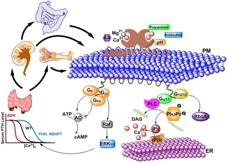
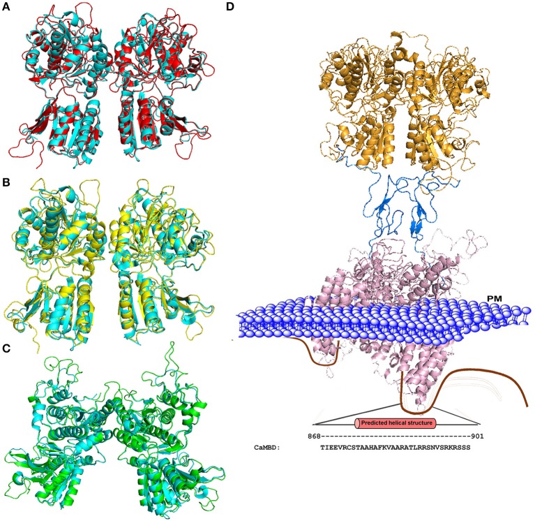
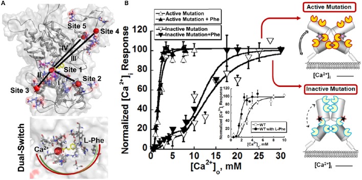
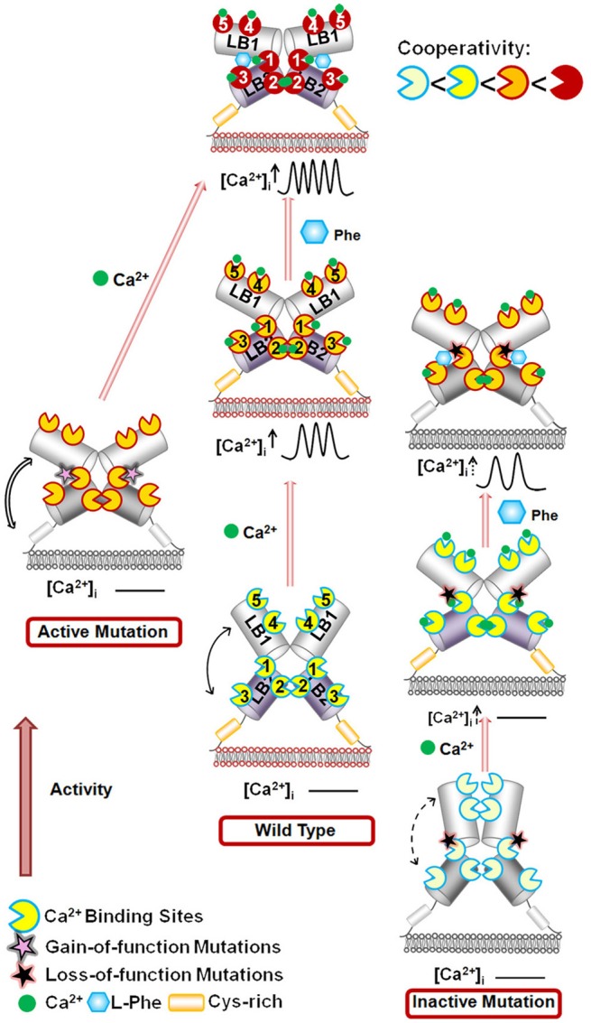
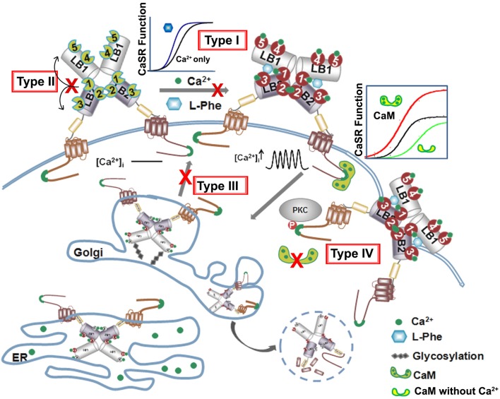
Ca2+-sensing receptors (CaSRs) play a central role in regulating extracellular calcium concentration ([Ca2+]o) homeostasis and many (patho)physiological processes in multiple organs. This regulation is orchestrated by a cooperative response to extracellular stimuli such as small changes in Ca2+, Mg2+, amino acids, and other ligands. In addition, CaSR is a pleiotropic receptor regulating several intracellular signaling pathways, including calcium mobilization and intracellular calcium oscillation. Nearly 200 mutations and polymorphisms have been found in CaSR in relation to a variety of human disorders associated with abnormal Ca2+ homeostasis. In this review, we summarize efforts directed at identifying binding sites for calcium and amino acids. Both homotropic cooperativity among multiple calcium binding sites and heterotropic cooperativity between calcium and amino acid were revealed using computational modeling, predictions, and site-directed mutagenesis coupled with functional assays. The hinge region of the bilobed Venus flytrap (VFT) domain of CaSR plays a pivotal role in coordinating multiple extracellular stimuli, leading to cooperative responses from the receptor. We further highlight the extensive number of disease-associated mutations that have also been shown to affect CaSR's cooperative action via several types of mechanisms. These results provide insights into the molecular bases of the structure and functional cooperativity of this receptor and other members of family C of the G protein-coupled receptors (cGPCRs) in health and disease states, and may assist in the prospective development of novel receptor-based therapeutics.

Keywords: amino acids; calcium sensing receptor; cooperativity; disease mutations; structure; trafficking.


Figures

Figure 1
Figure 1
Various types of agonists, including cations, peptides, amino acids, antibiotics, etc., can act on the extracellular calcium-sensing receptor (CaSR) to generate a complex intracellular signaling network. CaSR is also a pleiotropic receptor in its regulation of four G protein-mediated intracellular signaling pathways (Gq∕11, Gi∕o, Gs, and G12∕13). The correlations and crosstalk among different signaling cascades contribute the cooperative responses of intracellular calcium responses as well as parathyroid hormone (PTH) secretion (left) and intracellular calcium responses (right) to extracellular calcium. Bottom left: The sigmoidal relationship between calcium concentration in blood and PTH level in serum is demonstrated. Higher Ca2+ concentration is required for normal level of PTH in patients with familial hypocalciuric hypercalcemia (FHH) or patients with Neonatal Severe Primary Hyperparathyroidism (NSHPT), as the response curve shifts to the right; on the other hand, lower Ca2+ concentration than normal is enough to trigger PTH secretion in patients with autosomal dominant hypocalcemia (ADH). PM, Plasma membrane; ER, Endoplasmic reticulum; AA, arachidonic acid; AC, adenylate cyclase; cAMP, cyclic AMP; DAG, diacylglycerol; ERK1∕2, extracellular-signal-regulated kinase; Gs, Gi∕o,G12∕13, and Gq∕11, subunits of the s-, i-, 12/13, and q-type alpha subunit of heterotrimeric G proteins, respectively; Gβγ, G beta and gamma complex; IP3, inositol-1,4,5-trisphosphate; IP3R, inositol-1,4,5-trisphosphate receptor; PLC, phospholipase C; PI(4,5)P2, phoshatidylinositol-4,5-bisphosphate; RhoA, Ras homolog gene family, member A.
Figure 2
Figure 2
Homology models of the CaSR ECD. (A) Structural alignment of the mGluR1 holo form (cyan; PDB ID 1EWK, mGluR1 with glutamate) with the modeled structure of CaSR (red). (B) Structural alignment of mGluR1 apo form I (cyan, PDB ID 1EWV) with the modeled structure of CaSR (yellow). (C) Structural alignment of mGluR1 apo form II (cyan, PDB ID 1EWT) with the modeled structure of CaSR (green). (D) Homology model of the full CaSR structure. The ECD is based on the crystal structure of CaSR, the cysteine rich domain modeled from mGluR3 (PDB ID 2E4U), and the 7TM domain is modeled from mGluR1 with a ligand (PDB ID 4OR2). The C-terminal of CaSR can interact with protein kinases, ubiquitin ligase, CaM, etc. The region including residues from 868 to 901 is predicted to be CaM binding site. Mutations on the CaM binding site compromise the stability of surface expressed CaSR (Huang et al., 2010). PM: Plasma Membrane.
Figure 3
Figure 3
Molecular connectivity and heterotrophic cooperativity. (A) The correlated motions between calcium binding site 1 and the other calcium binding sites, designated by roman numbers (I~V), are mapped onto the CaSR ECD models. The correlation map and parameters for molecular dynamics simulation can be found in the paper (Zhang et al., 2014a). Bottom: The hetero-communication between Ca2+ and an amino acid functions as a dual switch that enhances the function of CaSR by positively impacting multiple Ca2+-binding sites within the ECD. Red sphere: Ca2+. Two headed arrow: both ligands can affect the binding of the other, therefore, the downstream signaling pathways. (B) The intracellular Ca2+ responses of HEK293 cells transiently overexpressing an active mutation or an inactive mutation. Inset: Representative response of WT CaSR to the changes of extracellular Ca2+ in the absence or presence of L-Phe, showing homotropic, and heterotropic cooperativity. Some disease mutations can interrupt the homotropic cooperativity shown as intracellular calcium responses to changes in the extracellular calcium concentration as well as the heterotropic cooperativity in the presence of allosteric modulators (e.g., L-Phe). Open circles: in the absence of L-Phe; Closed circles: in the presence of 5 mM L-Phe. Yellow “pacman”: Ca2+ binding sites in an active receptor; Blue “pacman”: Ca2+ binding sites in an inactive receptor. Pink stars: gain-of-function mutations in the hinge region. Black stars: loss-of-function mutations in the hinge region. Two headed arrows with solid lines: enhanced correlation motions between different Ca2+ binding sites. Two headed arrows with dash lines: impaired correlation motions.
Figure 4
Figure 4
Schematic representation of the mechanisms underlying the effects of the mutations on the CaSR and the modulation of receptor activity by extracellular Ca2+ and L-Phe. Ca2+ and L-Phe modulate the activity as well as the cooperativity of CaSR (the color changes of the receptor from ivory to red indicate an increase in functional activity). Elevating [Ca2+]o, e.g., to 3.0 mM, is proposed to change the basal WT CaSR status into an active form in a positive homotropic cooperative manner and further trigger intracellular Ca2+ oscillations. L-Phe binds to the hinge region between lobe 1 and lobe 2, modulating the receptor together with Ca2+ in a positive heterotropic cooperative way. This could potentiate conversion of the receptor to a “fully active” form associated with a higher frequency of intracellular Ca2+ oscillations and a left-shifted EC50. Loss-of-function CaSR mutants (indicated by ivory color) could cause a disruption of the cooperativity among the various Ca2+-binding sites as shown by impaired correlation motions (dashed arrows). The impaired receptor function and the cross-talk between Ca2+-binding sites can be at least be partially rescued for some mutants by L-Phe (e.g., P221Q). However, if the mutation interferes with the interaction between CaSR and L-Phe, the function of the receptor may not be fully recovered (e.g., L173P). CaSR gain-of-function mutants (left) exhibit enhanced correlated motions (double line arrows) and their activity is not further potentiated by L-Phe, potentially due to a ceiling effect. LB1(2): Lobe 1 or lobe 2 in the VFT domain of CaSR.
Figure 5
Figure 5
Summary of four types of disease mutations. The CaSR trafficking, expression and surface stabilization contribute to its functional cooperativity. Type I disease associated mutations directly alter key calcium and ligand binding capability at or in close proximity of the predicted ligand binding sites in the ECD. Type II mutations alter CaSR function especially EC50 or Hill number without altering surface expression or trafficking, but affect the molecular connectivity between different ligand binding sites. Type III mutations disrupt the cooperativity via interfering with the receptor cell surface expression and Type IV mutations largely affect potency of cooperativity by altering trafficking and protein stability via interaction with proteins binding to the CaSR intracellular C-tail.

References

    1. Alexander S. T., Hunter T., Walter S., Dong J., Maclean D., Baruch A., et al. (2015). Critical cysteine residues in both the calcium-sensing receptor and the allosteric activator AMG 416 underlie the mechanism of action. Mol. Pharmacol. 88, 853–865. 10.1124/mol.115.098392 - DOI - PubMed
    1. Alfadda T. I., Saleh A. M., Houillier P., Geibel J. P. (2014). Calcium-sensing receptor 20 years later. Am. J. Physiol. Cell Physiol. 307, C221–C231. 10.1152/ajpcell.00139.2014 - DOI - PMC - PubMed
    1. Arulpragasam A., Magno A. L., Ingley E., Brown S. J., Conigrave A. D., Ratajczak T., et al. (2012). The adaptor protein 14-3-3 binds to the calcium-sensing receptor and attenuates receptor-mediated Rho kinase signalling. Biochem. J. 441, 995–1006. 10.1042/BJ20111277 - DOI - PubMed
    1. Awata H., Huang C., Handlogten M. E., Miller R. T. (2001). Interaction of the calcium-sensing receptor and filamin, a potential scaffolding protein. J. Biol. Chem. 276, 34871–34879. 10.1074/jbc.M100775200 - DOI - PubMed
    1. Bai M. (1999). Structure and function of the extracellular calcium-sensing receptor (Review). Int. J. Mol. Med. 4, 115–125. 10.3892/ijmm.4.2.115 - DOI - PubMed

LinkOut - more resources