Review
. 2015 Oct 5;6(1):198-207.
doi: 10.1515/tnsci-2015-0021. eCollection 2015.

Arousal, motor control, and parkinson's disease

Affiliations
Review

Arousal, motor control, and parkinson's disease

E Garcia-Rill et al. Transl Neurosci. .

Abstract

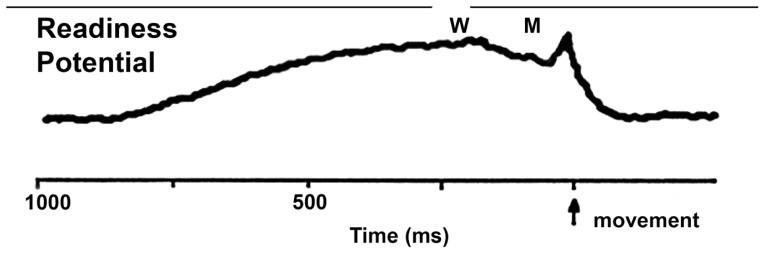
This review highlights the most important discovery in the reticular activating system (RAS) in the last 10 years, the manifestation of gamma (γ) band activity in cells of the RAS, especially in the pedunculopontine nucleus (PPN), which is in charge of the high frequency states of waking and rapid eye movement sleep. This discovery is critical to understanding the modulation of movement by the RAS and how it sets the background over which we generate voluntary and triggered movements. The presence of γ band activity in the RAS is proposed to participate in the process of preconscious awareness, and provide the essential stream of information for the formulation of many of our actions. Early findings using stimulation of this region to induce arousal, and also to elicit stepping, are placed in this context. This finding also helps explain the novel use of PPN deep brain stimulation for the treatment of Parkinson's disease, although considerable work remains to be done.

Keywords: Arousal; Calcium channels; Deep brain stimulation; Mu rhythm; P13 potential; P50 potential; Parkinson’s disease; Readiness potential.


Conflict of interest statement

The authors have no conflicts of interest.

Figures

Figure 1
Figure 1
The human readiness potential (RP). Representation of a vertex-recorded RP and the timing reported by subjects performing an uncued voluntary movement. The estimation of the sense of will (signified by “W”) or intent to move occurred well after the beginning of the RP, and the sensation of movement (signified by “M”) occurred even later, and well after the beginning of the RP as in the Libet study [11].
Figure 2
Figure 2
The pedunculopontine nucleus (PPN). A. Sagittal plane. Histochemical NADPH-diaphorase (reduced nicotinamide adenine dinucleotide phosphate diaphorase) labeling of only the cholinergic neurons in the PPN revealed the wedge-shaped structure of this nucleus. Posterodorsally is located the pars compacta of the PPN, which is located within the lateral cuneiform nucleus (LCN) ventral to the inferior colliculus (IC). As the nucleus descends, its cells are intermixed with the fibers of the superior cerebellar peduncle (SCP). The body or pars dissipata of the PPN is located more ventrally and extends to the posterior edge of the substantia nigra (SN). Anterior is to the right. B. Semi-horizontal. This section is angled along the long axis of the PPN from the posterodorsal to the medioventral direction (see Figure 2A for sagittal orientation). Histochemical NADPH-diaphorase labeling of cholinergic cells shows that PPN neurons of the pars compacta (pc) are located dorsally, are found within the cuneiform nucleus (CF), and laterally to the locus coeruleus (LC). Medial to the LC are cells of the laterodorsal tegmental nucleus (LDT) embedded within the central gray. Laterally, the pars dissipata (pd) of the PPN descends in an arc towards the posterior edge of the SN.
Figure 3
Figure 3
Pedunculopontine nucleus (PPN) neuronal properties. A. Increasing steps of current (increase of 30 pA per step, each step was 500 ms in duration, 2.5 s latency between each step, and the record was truncated between current steps and spliced to show only the current steps) caused cells to fire action potentials at higher frequencies until reaching a plateau at 40–60 Hz. This cell fired maximally at 54 Hz, which is within the γ frequency range [48]. B. Current clamp recording of a PPN neuron in the presence of fast synaptic blockers and tetrodotoxin (TTX), to which were applied current steps of increasing amplitude (dark gray record represents the response to lower amplitude square current while light gray record represents the response to higher amplitude square current steps). Note that the membrane potential failed to be maintained and repolarized below the window for high threshold, voltage-dependent calcium channels (−20 mV). C. Recordings in the same neuron but using ramps of increasing amplitude (dark gray record represents the response to lower amplitude current ramp while the light gray record represents the response to higher amplitude current ramp). Note that the membrane potential could be gradually increased to induce membrane oscillations that could be maintained within the window for activation of high threshold calcium channels (around −20 mV in the soma). Further studies showed that these oscillations were mediated by N- and P/Q-type voltage-dependent calcium channels [49].

References

    1. Llinas RR. I of the Vortex; from neurons to self. MIT Press; Cambridge, MA, USA: 2001.
    1. Eckhorn R, Bauer R, Jordan W, Brosch M, Kruse W, Munk M, et al. Coherent oscillations: a mechanism of feature linking in the visual system? Biol Cybern. 1988;60:121–130. - PubMed
    1. Gray CM, Singer W. Stimulus-specific neuronal oscillations in orientation columns of cat visual cortex. Proc Natl Acad Sci USA. 1989;86:1698–1702. - PMC - PubMed
    1. Llinas RR, Ribary U, Contreras D, Pedroarena C. The neuronal basis for consciousness. Philos Trans R Soc Lond B Biol Sci. 1998;353:1841–1849. - PMC - PubMed
    1. Pedroarena C, Llinás RR. Dendritic calcium conductances generate high-frequency oscillation in thalamocortical neurons. Proc Natl Acad Sci USA. 1997;94:724–728. - PMC - PubMed