

Review
doi: 10.1259/bjr.20160348.
Epub 2016 Oct 17.
Simple diagrammatic method to delineate male urethra in prostate cancer radiotherapy: an MRI based approach
Affiliations
- PMID: 27748126
- PMCID: PMC5604912
- DOI: 10.1259/bjr.20160348
Item in Clipboard
Review
Simple diagrammatic method to delineate male urethra in prostate cancer radiotherapy: an MRI based approach
Br J Radiol.
2016 Dec.
Abstract
Stereotactic body radiotherapy (SBRT) is being increasingly utilized in the treatment of prostate cancer. With the advent of high-precision radiosurgery systems, it is possible to obtain dose distributions akin to high-dose rate brachytherapy with SBRT. However, urethral toxicity has a significant impact on the quality of life in patients with prostate cancer. Contouring the male urethra on a CT scan is difficult in the absence of an indwelling catheter. In this pictorial essay, we have used the MRI obtained for radiotherapy planning to aid in the delineation of the male urethra and have attempted to define guidelines for the same.
Figures
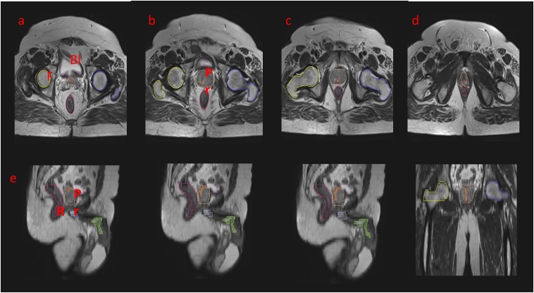
An axial T2 weighted sequence showing the
stepwise delineation of the prostatic urethra: the prostatic urethra
has been marked as a tubular structure in red–green. (a)
Identifying the bladder neck: the hyperintense signal is
representing the urine in the bladder. (b–d) It is traced
further into the parenchyma of the prostate. This is representing
the prostatic urethra. (e–g) The sagittal sectional
correlation to aid in delineation: the urethra as a whole has been
depicted. (g) The prostatic, bulbar and penile parts of the urethra
are shown. (h) A coronal section is illustrating the complete
prostatic urethra. Bl, bladder; F, femur; Pr, prostate; R,
rectum.
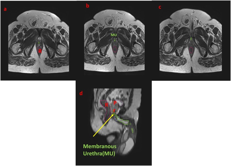
The axial T2 weighted sequence showing
the stepwise delineation of the prostatic urethra: the membranous
urethra (MU) has been marked as a tubular structure in green.
(a–c) Urogenital diaphragm: the MU is traversing through it
and ending at its inferior portion, beyond which it is continuing as
the bulbar part. (d) The coronal section with an arrow is denoting
the MU. Pr, prostate; R, rectum.

Axial T2 MRI: the penile bulb (corpus
spongiosum) has been identified and the bulbar urethra is starting
at the inferior border of the urogenital diaphragm. It is delineated
in green. (a) The penile bulb has been marked in purple, with the
bulbar urethra marked in green. (b, c) The bulbar urethra is
continuing as the penile urethra.
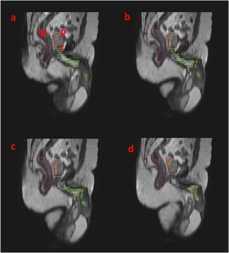
Sagittal T2 MRI: for correlating with the
axial sections, (a–d) the penile bulb has been marked in
purple. The bulbar urethra and penile urethra have been marked in
green. Bl, bladder; Pr, prostate.
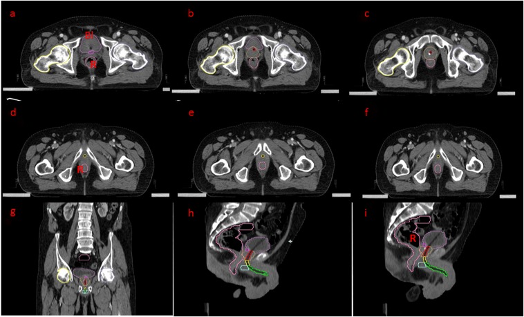
Axial and sagittal CT scans with catheter in situ:
(a–c) An axial CT scan—the bulb of Foley's
catheter is seen in the bladder. The bladder neck has been
delineated in pink, the prostate in yellow. The prostatic urethra is
traced along the catheter (red). (d–f) The prostatic urethra
is continuing as the membranous part, delineated in yellow. (g) The
coronal section is correlating the delineation. (h, i) Sagittal
sections for correlating the course of the urethra along
Foley's catheter. Bl, bladder; R, rectum.
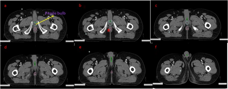
Axial CT scan with catheter in situ: (a–c)
the bulbar urethra has been contoured in blue. (d–f) The
penile urethra is traced till the meatus (green). R, rectum.
References
-
- Gray H, Clemente C. Anatomy of the human body. Philadelphia, PA: Lea & Febiger; 1985.
-
- Ryu J, Kim B. MR imaging of the male and female urethra. Radiographics 2001; 21: 1169–85. doi: https://doi.org/10.1148/radiographics.21.5.g01se121169 - DOI - PubMed
-
- Zietman AL, DeSilvio ML, Slater JD, Rossi CJ, Jr, Miller DW, Adams JA, et al. . Comparison of conventional-dose vs high-dose conformal radiation therapy in clinically localized adenocarcinoma of the prostate: a randomized controlled trial. JAMA 2005; 294: 1233–9. doi: https://doi.org/10.1001/jama.294.10.1233 - DOI - PubMed
-
- Viswanathan AN, Yorke ED, Marks LB, Eifel PJ, Shipley WU. Radiation dose-volume effects of the urinary bladder. Int J Radiat Oncol Biol Phys 2010; 76(Suppl. 3): S116–22. doi: https://doi.org/10.1016/j.ijrobp.2009.02.090 - DOI - PMC - PubMed
-
- Zelefsky MJ, Aschkenasy E, Kelsen S, Leibel SA. Tolerance and early outcome results of postprostatectomy three-dimensional conformal radiotherapy. Int J Radiat Oncol Biol Phys 1997; 39: 327–33. doi: https://doi.org/10.1016/S0360-3016(97)00056-4 - DOI - PubMed
Publication types
MeSH terms
LinkOut - more resources
Full Text Sources
Other Literature Sources
Medical
