

Sex hormone-binding globulin regulation of androgen bioactivity in vivo: validation of the free hormone hypothesis
- PMID: 27748448
- PMCID: PMC5066276
- DOI: 10.1038/srep35539
Sex hormone-binding globulin regulation of androgen bioactivity in vivo: validation of the free hormone hypothesis
Abstract
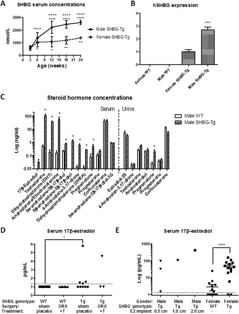
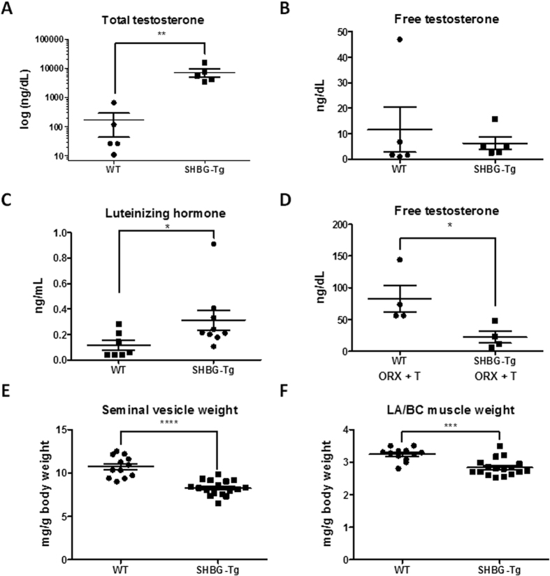
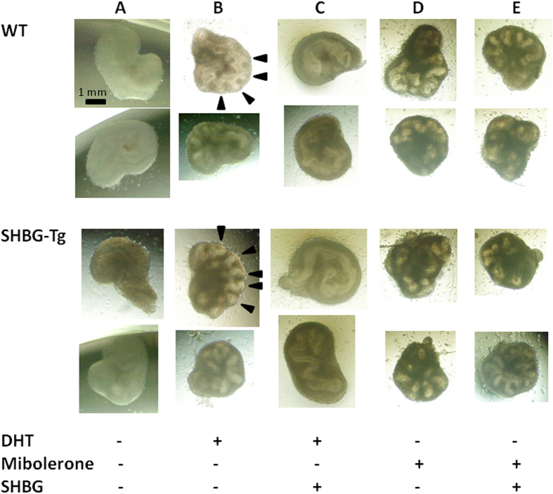
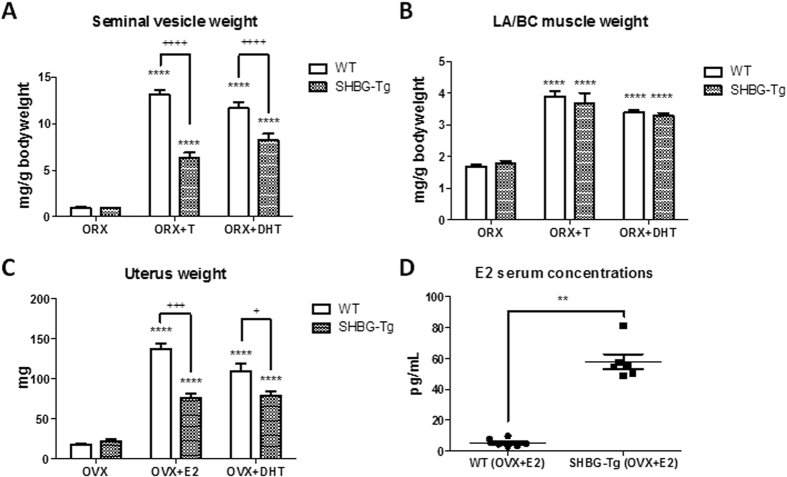
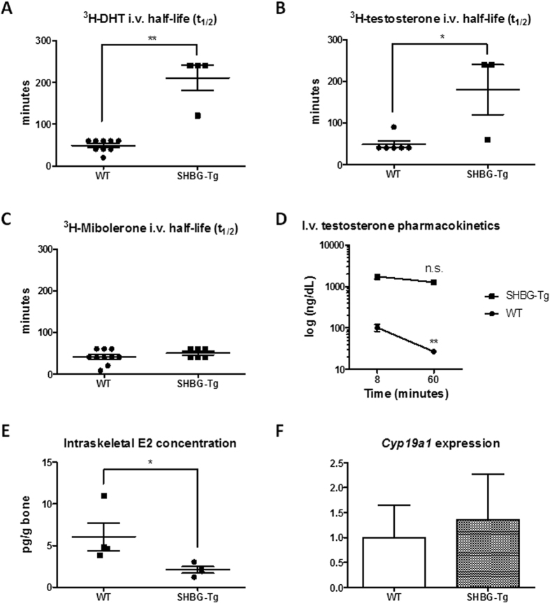
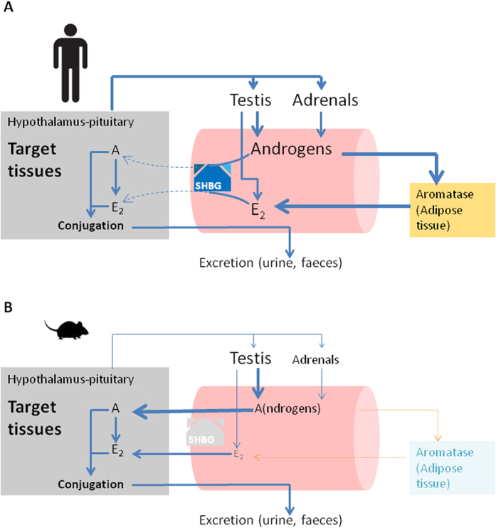
Sex hormone-binding globulin (SHBG) is the high-affinity binding protein for androgens and estrogens. According to the free hormone hypothesis, SHBG modulates the bioactivity of sex steroids by limiting their diffusion into target tissues. Still, the in vivo physiological role of circulating SHBG remains unclear, especially since mice and rats lack circulating SHBG post-natally. To test the free hormone hypothesis in vivo, we examined total and free sex steroid concentrations and bioactivity on target organs in mice expressing a human SHBG transgene. SHBG increased total androgen and estrogen concentrations via hypothalamic-pituitary feedback regulation and prolonged ligand half-life. Despite markedly raised total sex steroid concentrations, free testosterone was unaffected while sex steroid bioactivity on male and female reproductive organs was attenuated. This occurred via a ligand-dependent, genotype-independent mechanism according to in vitro seminal vesicle organ cultures. These results provide compelling support for the determination of free or bioavailable sex steroid concentrations in medicine, and clarify important comparative differences between translational mouse models and human endocrinology.
Figures
Similar articles
-
Effects of sex hormone-binding globulin (SHBG) on androgen bioactivity in vitro.Mol Cell Endocrinol. 2016 Dec 5;437:280-291. doi: 10.1016/j.mce.2016.08.041. Epub 2016 Aug 26. Mol Cell Endocrinol. 2016. PMID: 27576188
-
Molecular interactions between sex hormone-binding globulin and nonsteroidal ligands that enhance androgen activity.J Biol Chem. 2020 Jan 31;295(5):1202-1211. doi: 10.1074/jbc.RA119.011051. Epub 2019 Dec 18. J Biol Chem. 2020. PMID: 31852737 Free PMC article.
-
Selective cleavage of human sex hormone-binding globulin by kallikrein-related peptidases and effects on androgen action in LNCaP prostate cancer cells.Endocrinology. 2012 Jul;153(7):3179-89. doi: 10.1210/en.2012-1011. Epub 2012 Apr 30. Endocrinology. 2012. PMID: 22547569
-
Sex hormone-binding globulin is synthesized in target cells.J Endocrinol. 2002 Oct;175(1):113-20. doi: 10.1677/joe.0.1750113. J Endocrinol. 2002. PMID: 12379495 Review.
-
Structural analyses of sex hormone-binding globulin reveal novel ligands and function.Mol Cell Endocrinol. 2010 Mar 5;316(1):13-23. doi: 10.1016/j.mce.2009.09.005. Epub 2009 Sep 11. Mol Cell Endocrinol. 2010. PMID: 19748550 Review.
Cited by
-
Metabolic dysfunction-associated steatotic liver disease-associated fibrosis and cardiac dysfunction in patients with type 2 diabetes.World J Cardiol. 2024 Oct 26;16(10):580-594. doi: 10.4330/wjc.v16.i10.580. World J Cardiol. 2024. PMID: 39492975 Free PMC article.
-
Chronic liver diseases and erectile dysfunction.Front Public Health. 2023 Jan 6;10:1092353. doi: 10.3389/fpubh.2022.1092353. eCollection 2022. Front Public Health. 2023. PMID: 36684968 Free PMC article. Review.
-
Free Testosterone Reflects Metabolic as well as Ovarian Disturbances in Subfertile Oligomenorrheic Women.Int J Endocrinol. 2018 Sep 10;2018:7956951. doi: 10.1155/2018/7956951. eCollection 2018. Int J Endocrinol. 2018. PMID: 30275830 Free PMC article.
-
Skeletal muscle wasting: the estrogen side of sexual dimorphism.Am J Physiol Cell Physiol. 2022 Jan 1;322(1):C24-C37. doi: 10.1152/ajpcell.00333.2021. Epub 2021 Nov 17. Am J Physiol Cell Physiol. 2022. PMID: 34788147 Free PMC article. Review.
-
Dehydroepiandrosterone Supplementation Results in Varying Tissue-specific Levels of Dihydrotestosterone in Male Mice.Endocrinology. 2022 Oct 23;163(12):bqac163. doi: 10.1210/endocr/bqac163. Endocrinology. 2022. PMID: 36201601 Free PMC article.
References
-
- Mendel C. M. The free hormone hypothesis: a physiologically based mathematical model. Endocr Rev 10, 232–274 (1989). - PubMed
-
- Bhasin S. et al. Reference ranges for testosterone in men generated using liquid chromatography tandem mass spectrometry in a community-based sample of healthy nonobese young men in the Framingham Heart Study and applied to three geographically distinct cohorts. J Clin Endocrinol Metab 96, 2430–2439 (2011). - PMC - PubMed
-
- Rosner W., Auchus R. J., Azziz R., Sluss P. M. & Raff H. Position statement: Utility, limitations, and pitfalls in measuring testosterone: an Endocrine Society position statement. J Clin Endocrinol Metab 92, 405–413 (2007). - PubMed
Publication types
MeSH terms
Substances
LinkOut - more resources
Full Text Sources
Other Literature Sources
Miscellaneous
