. 2016 Oct 17:6:35416.
doi: 10.1038/srep35416.

Enhanced cryopreservation of MSCs in microfluidic bioreactor by regulated shear flow

Affiliations

Enhanced cryopreservation of MSCs in microfluidic bioreactor by regulated shear flow

Akalabya Bissoyi et al. Sci Rep. .

Abstract

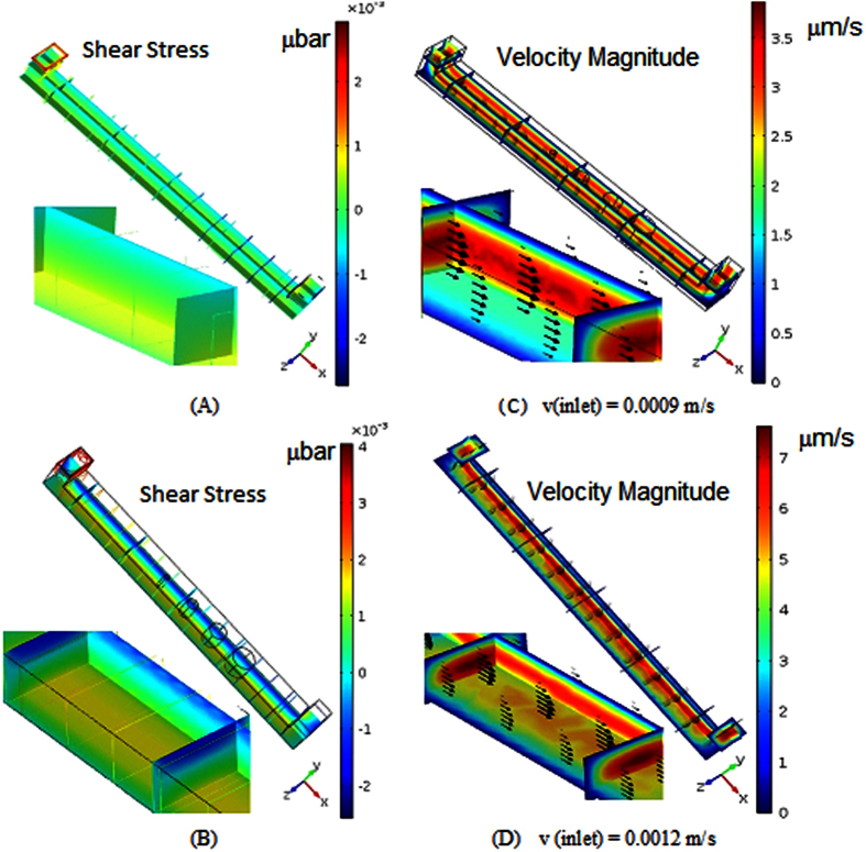
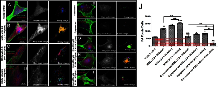
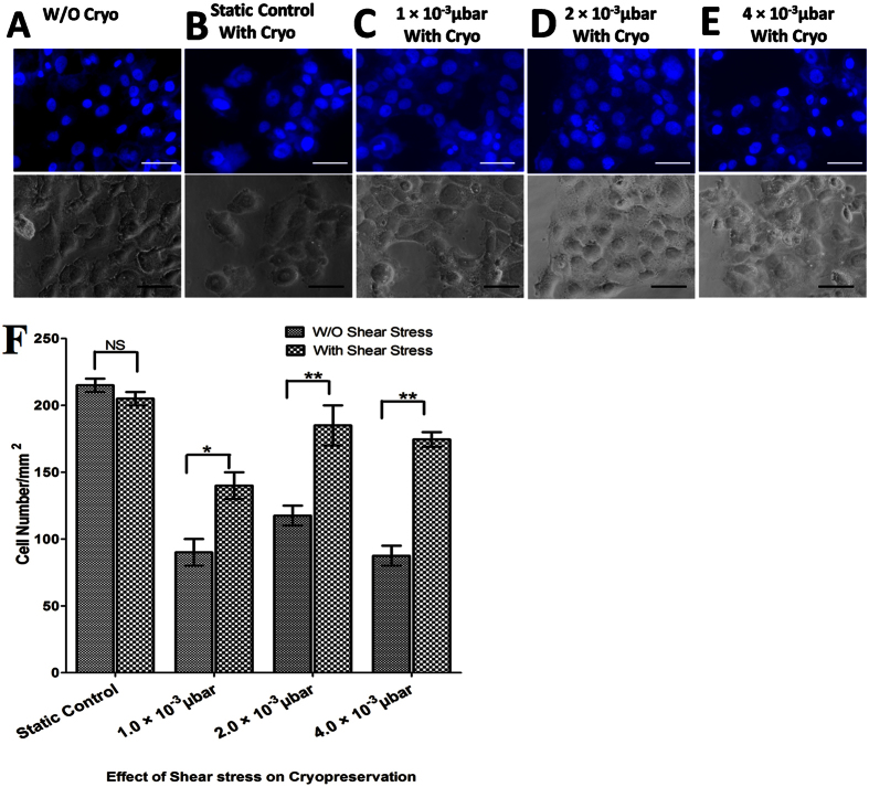
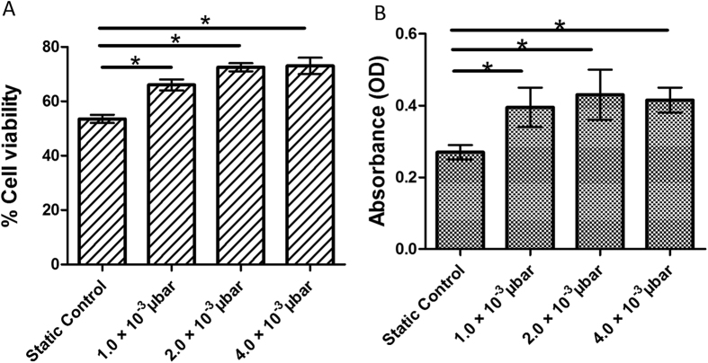
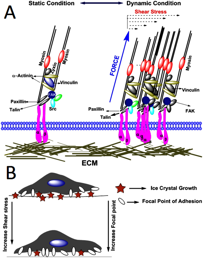
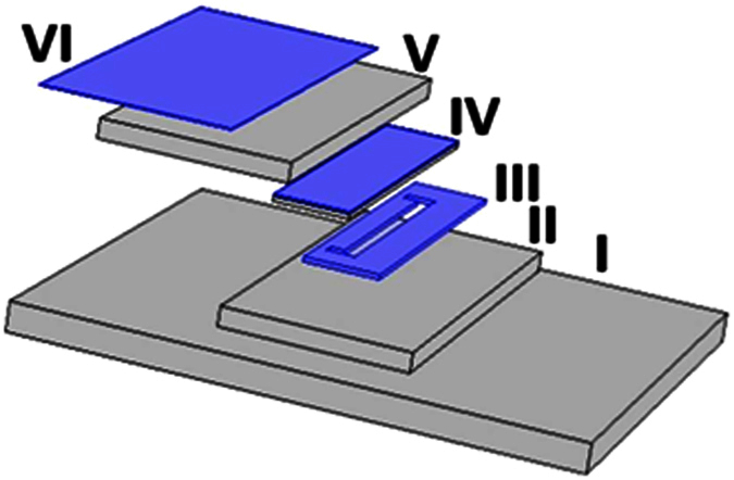
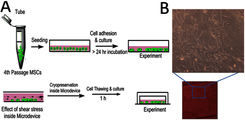
Cell-matrix systems can be stored for longer period of time by means of cryopreservation. Cell-matrix and cell-cell interaction has been found to be critical in a number of basic biological processes. Tissue structure maintenance, cell secretary activity, cellular migration, and cell-cell communication all exist because of the presence of cell interactions. This complex and co-ordinated interaction between cellular constituents, extracellular matrix and adjacent cells has been identified as a significant contributor in the overall co-ordination of tissue. The prime objective of this investigation is to evaluate the effects of shear-stress and cell-substrate interaction in successful recovery of adherent human mesenchymal-stem-cells (hMSCs). A customized microfluidic bioreactor has been used for the purpose. We have measured the changes in focal-point-adhesion (FPAs) by changing induced shear stress inside the bioreactor. The findings indicate that with increase in shear stress, FPAs increases between substrate and MSCs. Further, experimental results show that increased FPAs (4e-3 μbar) enhances the cellular survivability of adherent MSCs. Probably, for the first time involvement of focal point interaction in the outcome of cryopreservation of MSCs has been clarified, and it proved a potentially new approach for modification of cryopreservation protocol by up-regulating focal point of cells to improve its clinical application.


Conflict of interest statement

The authors declare no competing financial interests.

Figures

Figure 1
Figure 1
(A,B) Contour plot of Shear- stress distribution within micro-channel at different flow rates; (C,D) Contour plot of Velocity magnitude of cell-media composites in micro-channel at three different inlet flow rates.
Figure 2
Figure 2
Flow-cytometric analysis showing expression of positive Mesenchymal stem cells (MSC) markers CD44 (95%), CD73 (98%), CD90 (99%) and CD105 (96%) as well as negative MSC markers CD34 (1.2%), CD45 (2%) and CD14 (3.5%).
Figure 3
Figure 3. Vinculin associated focal point adhesions: Distribution of actin filaments and vinculin in human mesenchymal stem cells, showing cytoskeletal organization.
Actin filament, vinculin and nucleus are pseudocolor in green, red, and blue respectively. (A) 8th day, MSCs seeded in microchannel without any shear stress. (B) 8th day MSCs seeded in microchannel under 1 × 10−3 μbar. (C) 8th day MSCs seeded in microchannel under 2 × 10−3 μbar. (D) 8th day MSCs seeded in microchannel under 4 × 10−3 μbar. (E) Static Control (F) MSCs seeded in microfluidic device with cryopreservation treatment. Cryopreserved MSCs after 8 days of (G) 1 × 10−3 μbar, (H) 2 × 10−3 μbar, (I) 4 × 10−3 μbar shear stress inside microchannel. (J) Quantitative difference in focal points in MSCs at various shear stresses compare to static control. (NS: > 0.05, ** < 0.01, *** < 0.005).
Figure 4
Figure 4
(AE) Phase contrast and DAPI staining images of MSCs. MSCs were grown with or without shear stress. Representative images of (A) MSCs without cryopreservation, (B) MSCs with cryopreservation-static control. Cryopreserved MSCs with shear stress (C) 1 × 10−3 μbar (D) 2 × 10−3 μbar (E) 4 × 10−3 μbar. (F) bar plot image of cell number per mm2 (NS: > 0.05, ** < 0.01, *** < 0.005).
Figure 5
Figure 5
(A) Bar plot image of immediate post-thaw viability of MSCs inside microchannel measured by trypan blue. (B) Bar plat image of viable MSCs 24 h after post thaw, as assessed by the MTT assay. (NS: > 0.05, ** < 0.01, ***).
Figure 6
Figure 6. Representative images for differentiation potential of MSCs seeded inside microchannel.
Multilineage differentiation of shear induced cryopreserved MSCs showing differentiation potential towards both osteogenic and adipogenic lineages. Adipogenic oil droplet formation and osteogenic calcium nodule formation was determined by Oil Red O and alizarin red assays respectively.
Figure 7
Figure 7. Determination of stemness of MSCs seeded inside microchannel.
No significant higher expression level of stemness markers (NANOG, OCT-4, SOX-2 and REX-1) were observed in cryopreserved MSCs in various shear stress compared to fresh and static control.
Figure 8
Figure 8. Schematic representation of cell substrate interaction in presence of shear stress.
(A) With increase in shear stress focal adhesion point increases. (B) Schematic representation showing with increase in number of focal point decreases the intracellular space between cell-ECM, results in lesser ice formation in intracellular space.
Figure 9
Figure 9
(Schematic of Backside Diffused Light Lithography (BDLL): (I) represents the UV trans-illuminator from which UV light passes through (II) glass substrate and (III) pGS photo-mask (a mask on which the pattern of the micro-channel is drawn) to expose (IV) SU-8 substrate to produce micro-channel of exactly same pattern by regulating the amount of energy allowed to pass through the spatial region of pGS photo-mask. (V) Quartz Glass is used to cover SU-8 coated glass, whereas, (VI) a tinted film is used at the top of the set-up for preventing it from exposed on light.
Figure 10
Figure 10
Illustration of cryopreservation strategy used for MSCs inside microfluidic chamber (A) Schematic representation of cell seceding and subsequent cryopreservation protocol (B) Phase contrast image of MSCs inside microfluidic chamber and it enlarge image with 10X magnification obtained from confocal microscope.

References

    1. Chamberlain G., Fox J., Ashton B. & Middleton J. Concise review: mesenchymal stem cells: their phenotype, differentiation capacity, immunological features, and potential for homing. Stem cells 25(11), 2739–2749 (2007). - PubMed
    1. Jiang Y. et al.. Pluripotency of mesenchymal stem cells derived from adult marrow. Nature 418(6893), 41–49 (2002). - PubMed
    1. Bhakta G. et al.. Cryoreservation of alginate–fibrin beads involving bone marrow derived mesenchymal stromal cells by vitrification. Biomaterials 30(3), 336–343 (2009). - PubMed
    1. Meryman H. T. Cryopreservation of living cells: principles and practice. Transfusion 47(5), pp. 935–945 (2007). - PubMed
    1. Bagchi A., Woods E. J. & Critser J. K. Cryopreservation and vitrification: recent advances in fertility preservation technologies. Expert review of medical devices (2014). - PubMed

Publication types