Review
. 2016 Dec;38(6):1623-1641.
doi: 10.3892/ijmm.2016.2777. Epub 2016 Oct 17.

Cardiovascular diseases related to ionizing radiation: The risk of low-dose exposure (Review)

Affiliations
Review

Cardiovascular diseases related to ionizing radiation: The risk of low-dose exposure (Review)

Bjorn Baselet et al. Int J Mol Med. 2016 Dec.

Abstract

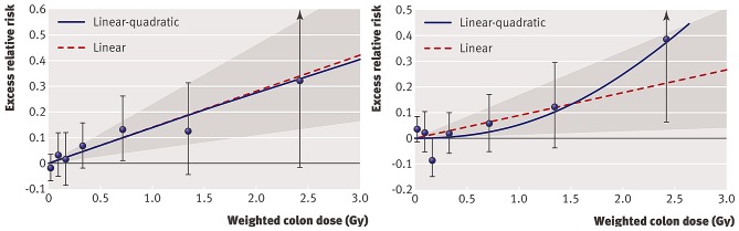
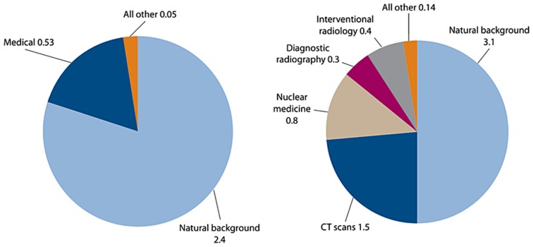
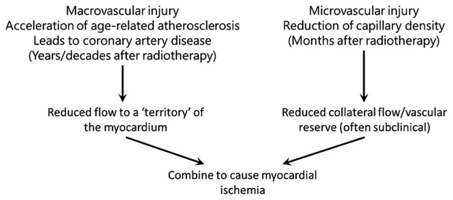
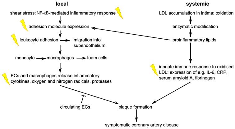
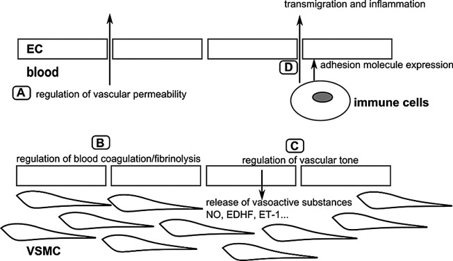
Traditionally, non-cancer diseases are not considered as health risks following exposure to low doses of ionizing radiation. Indeed, non-cancer diseases are classified as deterministic tissue reactions, which are characterized by a threshold dose. It is judged that below an absorbed dose of 100 mGy, no clinically relevant tissue damage occurs, forming the basis for the current radiation protection system concerning non-cancer effects. Recent epidemiological findings point, however, to an excess risk of non-cancer diseases following exposure to lower doses of ionizing radiation than was previously thought. The evidence is the most sound for cardiovascular disease (CVD) and cataract. Due to limited statistical power, the dose-risk relationship is undetermined below 0.5 Gy; however, if this relationship proves to be without a threshold, it may have considerable impact on current low‑dose health risk estimates. In this review, we describe the CVD risk related to low doses of ionizing radiation, the clinical manifestation and the pathology of radiation-induced CVD, as well as the importance of the endothelium models in CVD research as a way forward to complement the epidemiological data with the underlying biological and molecular mechanisms.


Figures

Figure 1
Figure 1
Radiation dose-response relationship (ERR/Gy) in the Life Span Study (LSS) cohort for death from stroke (left panel) and death from heart disease (right panel), showing linear-quadratic and linear functions. Shaded areas represent 95% confidence region for the fitted linear line. Error bars represent 95% CI for each dose category risks and the bullet represents the point estimate of risk for each dose category. The participants were divided into several dose categories according to their weighted colon dose (in Gy, γ dose plus 10 times neutron dose) (21).
Figure 2
Figure 2
Average annual effective dose/person received in 1980 (left panel) and 2006 (right panel) in the United States. The large increase in the use of ionizing radiation for medical purposes, in the period 1980–2006, contributed to a total increase from 3.0 mSv in 1980 to 6.2 mSv in 2006. Similar trends are observed in other industrialized countries (51).
Figure 3
Figure 3
Overview of the heart anatomy. (A) Illustration of the external anatomy with the major cardiac veins and arteries. (B) More detailed illustration of the pericardial sac that surrounds the heart.
Figure 4
Figure 4
Schematic overview of the development of an atherosclerotic lesion. In all steps, inflammation plays an important role. (A) A healthy artery with a well-functioning intact endothelium, a tunica intima, media and adventitia. VSMCs are mainly found in the tunica media but also in the tunica intima. (B) One of the initiating steps is the expression of adhesion molecules on the endothelium and the subsequent attraction of inflammatory blood cells (mainly monocytes). These monocytes will transmigrate to the intima where they will maturate to macrophages which will then transform to foam cells upon the uptake of ox-LDL. (C) Further progression to an atherosclerotic plaque includes the transmigration of VSMCs from the tunica media into the intima and the proliferation of VSMCs in the intima. There is also an enhanced production of extracellular matrix molecules, such as collagen, elastin and proteoglycans. Macrophages, foam cells and VSMCs can die, and released lipids will accumulate into the central region of the plaque, also denoted the lipid or necrotic core. (D) When a plaque ruptures it will induce thrombosis which is the major complication. The blood component will come in contact with the tissue factors present in the interior of the plaque triggering the formation of a thrombus which will hamper or even obstruct blood flow. The figure is based on a previous study (167). VSMCs, vascular smooth muscle cells; ox-LDL, oxidized low-density lipoprotein.
Figure 5
Figure 5
A theoretical overview of how radiation-induced macrovascular and microvascular pathologies can interact to cause myocardial ischemia, which may ultimately develop into clinical heart disease. The figure has been adapted from a previous study (5).
Figure 6
Figure 6
Overview of the major steps in the pathogenesis of coronary artery disease at the local and systemic level. Flashes indicate events that were also observed after radiation exposure, and which are mainly related to inflammation. ECs, endothelial cells; LDL, low-density lipoprotein; IL-6, interleukin-6; CRP, C-reactive protein. The figure has been adapted from a previous study (3).
Figure 7
Figure 7
Overview of the major physiological functions of the arterial endothelium. (A) The endothelium forms a selective barrier regulating the solute flux and fluid permeability between the blood and surrounding tissues (105). (B) The formation of a thrombus or blood clot is referred to as coagulation and the breakdown of a thrombus is referred to as fibrinolysis. Normal endothelium has anti-thrombotic and pro-fibrinolysis properties, and actively represses platelet adhesion and aggregation. Vessel damage or exposure to pro-inflammatory molecules will shift the balance towards more pro-thrombotic/anti-fibrinolysis actions (106,107). (C) To regulate vascular tone, the endothelium releases various vasodilatory factors such as NO and EDHF, or vasoconstrictive factors such as ET-1 which will modify VSMC function (108). (D) In the case of inflammation, endothelial permeability will be increased. Endothelial cells will also recruit immune cells via the expression of adhesion molecules, and mediate their transmigration towards the inner vascular wall (107). The figure is based on a previous study (103). ECs, endothelial cells; VSMCs, vascular smooth muscle cells; NO, nitric oxide; EDHF, endothelium-derived hyperpolarizing factor; ET-1, endothelin-1.
Figure 8
Figure 8
Electron structures of common ROS. Below each structure, its name and chemical formula are given. • represents an unpaired electron. ROS, reactive oxygen species.

References

    1. Stewart JR, Fajardo LF. Radiation-induced heart disease. Clinical and experimental aspects. Radiol Clin North Am. 1971;9:511–531. - PubMed
    1. Stewart FA. Mechanisms and dose-response relationships for radiation-induced cardiovascular disease. Ann ICRP. 2012;41:72–79. doi: 10.1016/j.icrp.2012.06.031. - DOI - PubMed
    1. Schultz-Hector S, Trott KR. Radiation-induced cardiovascular diseases: Is the epidemiologic evidence compatible with the radiobiologic data? Int J Radiat Oncol Biol Phys. 2007;67:10–18. doi: 10.1016/j.ijrobp.2006.08.071. - DOI - PubMed
    1. Clarke M, Collins R, Darby S, Davies C, Elphinstone P, Evans V, Godwin J, Gray R, Hicks C, James S, et al. Early Breast Cancer Trialists' Collaborative Group (EBCTCG) Effects of radiotherapy and of differences in the extent of surgery for early breast cancer on local recurrence and 15-year survival: An overview of the randomised trials. Lancet. 2005;366:2087–2106. doi: 10.1016/S0140-6736(05)67887-7. - DOI - PubMed
    1. Darby SC, Cutter DJ, Boerma M, Constine LS, Fajardo LF, Kodama K, Mabuchi K, Marks LB, Mettler FA, Pierce LJ, et al. Radiation-related heart disease: Current knowledge and future prospects. Int J Radiat Oncol Biol Phys. 2010;76:656–665. doi: 10.1016/j.ijrobp.2009.09.064. - DOI - PMC - PubMed