. 2016 Dec;13(12):1001-1004.
doi: 10.1038/nmeth.4033. Epub 2016 Oct 17.

Random-access scanning microscopy for 3D imaging in awake behaving animals

Affiliations

Random-access scanning microscopy for 3D imaging in awake behaving animals

K M Naga Srinivas Nadella et al. Nat Methods. 2016 Dec.

Abstract

Understanding how neural circuits process information requires rapid measurements of activity from identified neurons distributed in 3D space. Here we describe an acousto-optic lens two-photon microscope that performs high-speed focusing and line scanning within a volume spanning hundreds of micrometers. We demonstrate its random-access functionality by selectively imaging cerebellar interneurons sparsely distributed in 3D space and by simultaneously recording from the soma, proximal and distal dendrites of neocortical pyramidal cells in awake behaving mice.


Conflict of interest statement

Competing financial interests

The authors declare competing financial interests: details accompany the full text HTML version of the paper at http://www.nature.com/naturemethods/. Patents on the AOL technology have been filed (WO/2008/032061, WO/2011/131933).

Figures

Figure 1
Figure 1. Acousto-optic lens (AOL) two-photon microscope and random access multi-plane imaging across layers of the cerebellar cortex of an awake mouse.
a) Schematic diagram of the AOL 3D random access two-photon microscope configured for in vivo imaging. Inset box: Schematic diagram showing full Z-stack, multi-plane, sub-volume and patch scanning modes together with pointing. Imaging speed calculated for line-scanning and pointing mode using 50 ns and 4 μs dwell time, respectively. b) Theoretical relationship between AOL transmission efficiency and scan angle for different voxel dwell times when the AOL is focussed to 1 m (equivalent to Z=+135 μm with a 20x objective). c) AOL Z-stack (136 planes with 2 μm step at 100 ns dwell time) of GFP expressing inhibitory interneurons in mouse cerebellum. d) Averaged images of cerebellar interneurons expressing GCaMP6f in 4 planes acquired near-simultaneously at 19.5 Hz (100 ns/voxel), after post hoc movement correction. Coloured squares indicating the neuronal structures from which the fluorescent traces (right) were extracted. Background colours indicate layers in Supplementary Fig 5a. Grey trace at the bottom shows the speed of animal, with grey shading indicating periods of locomotion. Vertical bar indicates normalized change in fluorescence (ΔF/F) together with speed of locomotion (cm/s); and the horizontal scale bar indicates time (s).
Figure 2
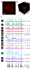
Figure 2. Random-access patch imaging of neurons in layer 2/3 of primary visual cortex in an awake behaving mouse.
a) Maximum intensity projection of red channel for cells in layer 2/3 of visual cortex sparsely expressing td-Tomato and GCaMP6f. b) Location of 14 X-Y patches within the imaging volume, distributed between 74 μm to 163 μm below the pia. c) Averaged images of cellular structures (green channel) scanned near-simultaneously at 50 ns/voxel in the 14 patches (51 x 50 voxels, 22 μm x 21.5 μm) after post hoc correction for brain movement. Traces to the right show ΔF/F responses extracted from each patch (52.8 Hz patch cycle rate, i.e. 1/(time to scan all patches)). Grey trace at the bottom shows the speed of the animal on the cylindrical treadmill. Blue-grey shading indicates periods of locomotion. Scale bars as defined in figure 1 legend.
Figure 3
Figure 3. Simultaneous dendritic and somatic imaging of pyramidal neurons in the visual cortex of awake mice.
a) Partially reconstructed image of a layer 5 neuron within the imaging volume (red box) together with the location of 3 user-selected patches intersecting the soma, apical dendrite and the nexus of the distal dendritic tuft. b) Averaged images of the soma and dendritic structures scanned near-simultaneously at 100 ns/voxel in the 3 patches (67 x 51 voxels, ~31 μm x ~23 μm) after post hoc correction for brain movement, together with mask outlines (green lines) for extraction of functional data. The acquisition rate was 209 Hz for the 3 patch cycle. Traces to the right show ΔF/F responses extracted from each colour coded patch while the animal was stationary. Dashed black box indicates measurements from the soma and dendrites that were confirmed to be from the same reconstructed cell, while the two other dendrites could not be unequivocally confirmed from the Z-stack image. Grey trace at the bottom shows the speed of the animal on the cylindrical treadmill. c) As for b but during locomotion. d) Two planes showing the somatic and dendritic sections of cells in layer 2/3 visual cortex sparsely expressing td-Tomato and GCaMP6f. e) Location of 3 sub-volumes within the imaging volume together with a partial reconstruction of an imaged L2/3 neuron that includes the soma, proximal dendrite and distal dendrite. Each sub-volume consists of 5-6 planes (224 x 59 voxels, 96 μm x 25 μm) 4 μm apart. f) Images of different regions of the pyramidal cell from the planes making up each sub-volume after post hoc movement correction. Traces show ΔF/F responses extracted from the cellular component present in each plane, where red, green and blue indicate soma, proximal dendrite and distal dendrite, respectively. Data were acquired at 50 ns/voxel with a sampling rate of 27.9 Hz for the 3 volume cycle. Grey trace at the bottom shows the speed of locomotion of the animal on the cylindrical wheel. Blue-grey shading indicates periods of locomotion. Scale bars as defined in figure 1 legend.

References

    1. Svoboda K, Yasuda R. Principles of two-photon excitation microscopy and its applications to neuroscience. Neuron. 2006;50:823–839. doi: 10.1016/j.neuron.2006.05.019. - DOI - PubMed
    1. Grewe BF, Voigt FF, van 't Hoff M, Helmchen F. Fast two-layer two-photon imaging of neuronal cell populations using an electrically tunable lens. Biomed Opt Express. 2011;2:2035–2046. doi: 10.1364/BOE.2.002035. - DOI - PMC - PubMed
    1. Kong L, et al. Continuous volumetric imaging via an optical phase-locked ultrasound lens. Nat Methods. 2015;12:759–762. doi: 10.1038/nmeth.3476. - DOI - PMC - PubMed
    1. Botcherby EJ, et al. Aberration-free three-dimensional multiphoton imaging of neuronal activity at kHz rates. Proc Natl Acad Sci U S A. 2012;109:2919–2924. doi: 10.1073/pnas.1111662109. - DOI - PMC - PubMed
    1. Kaplan A, Friedman N, Davidson N. Acousto-optic lens with very fast focus scanning. Opt Lett. 2001;26:1078–1080. - PubMed

MeSH terms

Substances