Comparative Study
. 2016 Dec;48(12):1570-1575.
doi: 10.1038/ng.3700. Epub 2016 Oct 17.

Prospective functional classification of all possible missense variants in PPARG

Affiliations
Comparative Study

Prospective functional classification of all possible missense variants in PPARG

Amit R Majithia et al. Nat Genet. 2016 Dec.

Abstract

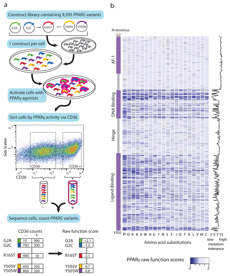
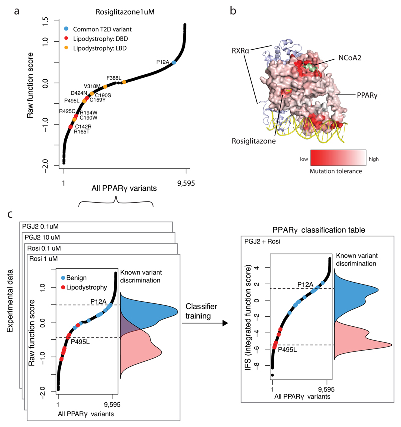
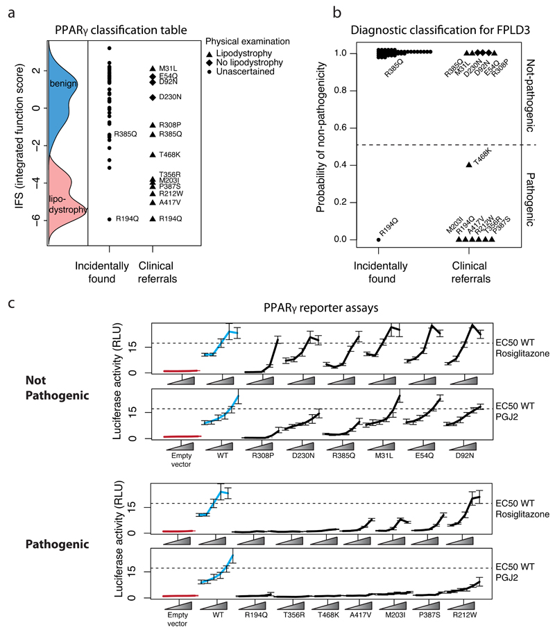
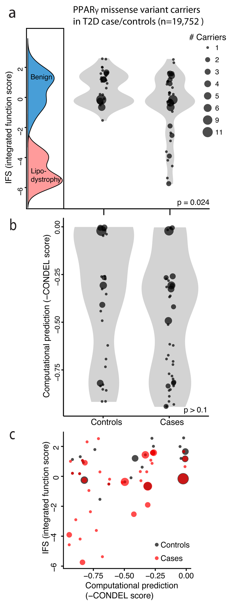
Clinical exome sequencing routinely identifies missense variants in disease-related genes, but functional characterization is rarely undertaken, leading to diagnostic uncertainty. For example, mutations in PPARG cause Mendelian lipodystrophy and increase risk of type 2 diabetes (T2D). Although approximately 1 in 500 people harbor missense variants in PPARG, most are of unknown consequence. To prospectively characterize PPARγ variants, we used highly parallel oligonucleotide synthesis to construct a library encoding all 9,595 possible single-amino acid substitutions. We developed a pooled functional assay in human macrophages, experimentally evaluated all protein variants, and used the experimental data to train a variant classifier by supervised machine learning. When applied to 55 new missense variants identified in population-based and clinical sequencing, the classifier annotated 6 variants as pathogenic; these were subsequently validated by single-variant assays. Saturation mutagenesis and prospective experimental characterization can support immediate diagnostic interpretation of newly discovered missense variants in disease-related genes.


Conflict of interest statement

No competing financial interests

Figures

Figure 1
Figure 1. Comprehensive functional testing of 9,595 PPARγ amino acid variants.
a) A library of 9,595 PPARG constructs was synthesized, each construct containing one amino acid substitution. The construct library was introduced into THP-1 monocytes (edited to lack the endogenous PPARG gene) such that each cell received a single construct. This polyclonal population of THP-1 monocytes was differentiated to macrophages and stimulated with PPARγ agonists (rosiglitazone, PGJ2); the stimulated macrophages were separated via fluoresence activated cell sorting according to expression of the PPARγ response gene CD36 into low (-) and high (+) activity bins. Each bin of cells was subject to next-generation sequencing at the transgenic PPARG locus to identify and tabulate introduced variants. PPARγ variant counts in the CD36 low and CD36 high bins were used to calculate a functional score for all 9,595 variants. b) Raw PPARγ function scores for each of the 9,595 variants plotted according to amino acid position along the PPARγ sequence. “Blue” denotes that any amino acid change away from reference results in low CD36 function score, whereas ”white” denotes that amino acid changes do not alter function; “grey” denotes the reference amino acid. Function scores summed by amino acid position are plotted to the right, denoting tolerance for any amino acid substitution away from reference.
Figure 2
Figure 2. Integrating experimental function to construct a PPARγ classification table.
a) Raw PPARγ function scores ranked for all 9,595 PPARγ variants tested. Highlighted in red are raw function scores of known lipodystrophy causing mutations if they reside in the DNA-binding domain (DBD) or in orange if they reside in the Ligand-binding domain (LBD). The common P12A variant is shown in blue. b) Mutation tolerance scores as described in Figure1 are shown color-coded and mapped onto the known crystal structure of PPARγ with RXRα, NCoA and Rosiglitazone. “Red” denotes that amino acid changes away from reference results in low CD36 function score, whereas ”white” denotes that amino acid changes do not alter function. c) Raw PPARγ function scores were obtained for 9,595 variants under four experimental conditions: 1) 1 μM Rosiglitazone, 2) 0.1 μM Rosiglitazone, 3) 10 μM Prostaglandin J2, and 4) 0.1 μM Prostaglandin J2. The function of known benign (n=13) and lipodystrophy-causing (n=11) variants are highlighted in blue and red respectively with their overall distributions overlaid. The raw function scores were combined into an integrated function score (IFS) after classifier training using linear discriminant analysis (LDA).
Figure 3
Figure 3. Experimental and clinical classification of novel missense PPARG variants identified in sequenced individuals.
a) Variants identified in patients plotted according to their integrated function score (IFS) alongside the IFS distributions of known benign, and lipodystrophy associated variants. b) Diagnostic classification for Familial Partial Lipodystrophy 3 (FPLD3) expressed as posterior probability of non-pathogenicity of PPARG variants shown in (a). Posterior probability was calculated by combining IFS with prevalence of lipodystrophy in the general population (1:100,000) or from patients referred for lipodystrophy/familial diabetes (1:7). c) The variants identified in patients were individually recreated and tested for their ability to activate luciferase reporter constructs containing three, tandemly-repeated, copies of the PPRE from the Acyl-CoA oxidase gene linked to the thymidine kinase promoter under varying doses of pharmacologic (rosiglitazone) or endogenous (prostaglandin J2; PGJ2) ligands (mean +/- S.E.M n =5). Variants are grouped according to not-pathogenic/pathogenic designation in (b).
Figure 4
Figure 4. Ability of PPARγ p.R212W to transactivate gene expression and bind DNA at endogenous enhancers
a) Ability of PPARγ2 WT or R212W mutant to activate luciferase reporter constructs containing FABP4 promoter under varying doses of pharmacologic (rosiglitazone 0-1μM) or endogenous (prostaglandin J2; PGJ2 0-10μM) ligands (mean +/- S.E.M n = 5). b) Comparison of the DNA binding properties of in vitro translated wild type or mutant PPARγ proteins, tested in electrophoretic mobility shift assays using either γ1 (R184W) or γ2 (R212W) mutants and radiolabelled PPREs from the acyl coenzyme A oxidase (AcCoA: 5’ ggaccAGGACAaAGGTCAcgtt 3’ ), fatty acid binding protein 4 (FABP4: 5’aaacaCAGGCAaAGGTCAgagg 3’) or muscle carnitine palmitoyl transferase 1 (CPT1: 5’ atcggTGACCTtTTCCCTaca 3’) promoters with retinoid X receptor (RXR) and increasing concentrations of ligand (Rosiglitazone 0 to 10uM). RL, reticulocyte lysate. c) PPARγ colored by mutation tolerance scores obtained under stimulation with 1μM Rosiglitazone in THP-1 cells. As in Figure 2b, red represents sites that exhibited low CD36 response when mutated away from WT. Arginine 212 is highlighted which occurs in the ‘hinge’ region of PPARγ connecting the DNA binding and ligand binding domains. The positively charged arginine side chain extends into the minor groove of DNA forming multiple hydrogen bonds with bases.
Figure 5
Figure 5. Relationship of PPARγ function to T2D risk in the general population.
a) Missense PPARγ variants identified from 19,752 sequenced type 2 diabetes (T2D) case/controls plotted according to IFS (integrated functional score) from the PPARγ classification table alongside the IFS distributions of known benign, and lipodystrophy associated variants. Each point represents a missense variant; point size denote the number of individuals carrying that variant. Among the 118 individuals carrying missense PPARγ variants T2D cases contained a long tail of low-functioning missense variants, which was notably absent from the distribution of variants observed in T2D controls (p = 0.024 two-sample Kolmogorov-Smirnov test). b) When the same 118 individuals were plotted according to computational prediction of deleteriousness no difference is distributions of functional variants is seen among T2D cases vs controls (p > 0.1 two-sample Kolmogorov-Smirnov test). c) Scatterplot of IFS vs computational prediction scores for PPARγ missense variants from T2D case/controls as described above.

References

    1. Majewski J, Schwartzentruber J, Lalonde E, Montpetit A, Jabado N. What can exome sequencing do for you? J Med Genet. 2011;48:580–9. - PubMed
    1. Gahl WA, et al. The National Institutes of Health Undiagnosed Diseases Program: insights into rare diseases. Genet Med. 2012;14:51–9. - PMC - PubMed
    1. Barroso I, et al. Dominant negative mutations in human PPARgamma associated with severe insulin resistance, diabetes mellitus and hypertension. Nature. 1999;402:880–3. - PubMed
    1. Jeninga E, Gurnell M. Functional implications of genetic variation in human PPAR [gamma] Trends in Endocrinology & …. 2009 - PubMed
    1. Majithia AR, et al. Rare variants in PPARG with decreased activity in adipocyte differentiation are associated with increased risk of type 2 diabetes. Proc Natl Acad Sci U S A. 2014;111:13127–32. - PMC - PubMed

Publication types