Modulation of topoisomerase IIα expression and chemosensitivity through targeted inhibition of NF-Y:DNA binding by a diamino p-anisyl-benzimidazole (Hx) polyamide
- PMID: 27750031
- PMCID: PMC5757371
- DOI: 10.1016/j.bbagrm.2016.10.005
Modulation of topoisomerase IIα expression and chemosensitivity through targeted inhibition of NF-Y:DNA binding by a diamino p-anisyl-benzimidazole (Hx) polyamide
Abstract
Background: Sequence specific polyamide HxIP 1, targeted to the inverted CCAAT Box 2 (ICB2) on the topoisomerase IIα (topo IIα) promoter can inhibit NF-Y binding, re-induce gene expression and increase sensitivity to etoposide. To enhance biological activity, diamino-containing derivatives (HxI*P 2 and HxIP* 3) were synthesised incorporating an alkyl amino group at the N1-heterocyclic position of the imidazole/pyrrole.
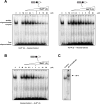
Methods: DNase I footprinting was used to evaluate DNA binding of the diamino Hx-polyamides, and their ability to disrupt the NF-Y:ICB2 interaction assessed using EMSAs. Topo IIα mRNA (RT-PCR) and protein (Immunoblotting) levels were measured following 18h polyamide treatment of confluent A549 cells. γH2AX was used as a marker for etoposide-induced DNA damage after pre-treatment with HxIP* 3 and cell viability was measured using Cell-Titer Glo®.
Results: Introduction of the N1-alkyl amino group reduced selectivity for the target sequence 5'-TACGAT-3' on the topo IIα promoter, but increased DNA binding affinity. Confocal microscopy revealed both fluorescent diamino polyamides localised in the nucleus, yet HxI*P 2 was unable to disrupt the NF-Y:ICB2 interaction and showed no effect against the downregulation of topo IIα. In contrast, inhibition of NF-Y binding by HxIP* 3 stimulated dose-dependent (0.1-2μM) re-induction of topo IIα and potentiated cytotoxicity of topo II poisons by enhancing DNA damage.
Conclusions: Polyamide functionalisation at the N1-position offers a design strategy to improve drug-like properties. Dicationic HxIP* 3 increased topo IIα expression and chemosensitivity to topo II-targeting agents.
General significance: Pharmacological modulation of topo IIα expression has the potential to enhance cellular sensitivity to clinically-used anticancer therapeutics. This article is part of a Special Issue entitled: Nuclear Factor Y in Development and Disease, edited by Prof. Roberto Mantovani.
Keywords: Chemosensitisation; DNA-binding polyamides; Gene modulation; NF-Y; Sequence selectivity; Topoisomerase IIα (topo IIα); Transcription factor-DNA interactions.
Copyright © 2016. Published by Elsevier B.V.
Figures


Similar articles
-
Modulation of topoisomerase IIalpha expression by a DNA sequence-specific polyamide.Mol Cancer Ther. 2007 Jan;6(1):346-54. doi: 10.1158/1535-7163.MCT-06-0503. Mol Cancer Ther. 2007. PMID: 17237293
-
Nuclear Localization and Gene Expression Modulation by a Fluorescent Sequence-Selective p-Anisyl-benzimidazolecarboxamido Imidazole-Pyrrole Polyamide.Chem Biol. 2015 Jul 23;22(7):862-75. doi: 10.1016/j.chembiol.2015.06.005. Epub 2015 Jun 25. Chem Biol. 2015. PMID: 26119998
-
Targeting the inverted CCAAT box 2 in the topoisomerase IIalpha promoter by JH-37, an imidazole-pyrrole polyamide hairpin: design, synthesis, molecular biology, and biophysical studies.Biochemistry. 2004 Sep 28;43(38):12249-57. doi: 10.1021/bi048785z. Biochemistry. 2004. PMID: 15379563
-
Nuclear factor-Y binding to the topoisomerase IIalpha promoter is inhibited by both the p53 tumor suppressor and anticancer drugs.Mol Pharmacol. 2003 Feb;63(2):359-67. doi: 10.1124/mol.63.2.359. Mol Pharmacol. 2003. PMID: 12527807
-
The CCAAT-binding complex (CBC) in Aspergillus species.Biochim Biophys Acta Gene Regul Mech. 2017 May;1860(5):560-570. doi: 10.1016/j.bbagrm.2016.11.008. Epub 2016 Dec 8. Biochim Biophys Acta Gene Regul Mech. 2017. PMID: 27939757 Review.
Cited by
-
DNA-Binding Properties of New Fluorescent AzaHx Amides: Methoxypyridylazabenzimidazolepyrroleimidazole/pyrrole.Chembiochem. 2018 Sep 17;19(18):1979-1987. doi: 10.1002/cbic.201800273. Epub 2018 Aug 15. Chembiochem. 2018. PMID: 29974647 Free PMC article.
-
Bioactive pyrrole-based compounds with target selectivity.Eur J Med Chem. 2020 Dec 15;208:112783. doi: 10.1016/j.ejmech.2020.112783. Epub 2020 Aug 29. Eur J Med Chem. 2020. PMID: 32916311 Free PMC article. Review.
-
Structural Basis of Inhibition of the Pioneer Transcription Factor NF-Y by Suramin.Cells. 2020 Oct 29;9(11):2370. doi: 10.3390/cells9112370. Cells. 2020. PMID: 33138093 Free PMC article.
-
A survey of recent unusual high-resolution DNA structures provoked by mismatches, repeats and ligand binding.Nucleic Acids Res. 2018 Jul 27;46(13):6416-6434. doi: 10.1093/nar/gky561. Nucleic Acids Res. 2018. PMID: 29945186 Free PMC article. Review.
-
Targeting Transcription Factors for Cancer Treatment.Molecules. 2018 Jun 19;23(6):1479. doi: 10.3390/molecules23061479. Molecules. 2018. PMID: 29921764 Free PMC article. Review.
References
-
- White S, Baird EE, Dervan PB. On the pairing rules for recognition in the minor groove of DNA by pyrrole-imidazole polyamides. Chemistry & Biology. 1997;4:569–578. - PubMed
-
- Kielkopf CL, Baird EE, Dervan PB, Rees DC. Structural basis for G•C recognition in the DNA minor groove. Nature Structural Biology. 1998;5:104–109. - PubMed
-
- Dervan PB. Molecular recognition of DNA by small molecules. Bioorganic & medicinal chemistry. 2001;9:2215–2235. - PubMed
-
- Dervan PB, Edelson BS. Recognition of the DNA minor groove by pyrrole-imidazole polyamides. Current Opinion in Structural Biology. 2003:284–299. - PubMed
-
- Kageyama Y, Sugiyama H, Ayame H, Iwai A, Fujii Y, Huang LE, Kizaka-Kondoh S, Hiraoka M, Kihara K. Suppression of VEGF transcription in renal cell carcinoma cells by pyrrole-imidazole hairpin polyamides targeting the hypoxia responsive element. Acta oncologica (Stockholm, Sweden) 2006;45:317–324. - PubMed
Publication types
MeSH terms
Substances
Grants and funding
LinkOut - more resources
Full Text Sources
Other Literature Sources
Molecular Biology Databases

