. 2016 Dec;186(12):3225-3235.
doi: 10.1016/j.ajpath.2016.08.020. Epub 2016 Oct 15.

Hypophysitis Secondary to Cytotoxic T-Lymphocyte-Associated Protein 4 Blockade: Insights into Pathogenesis from an Autopsy Series

Affiliations

Hypophysitis Secondary to Cytotoxic T-Lymphocyte-Associated Protein 4 Blockade: Insights into Pathogenesis from an Autopsy Series

Patrizio Caturegli et al. Am J Pathol. 2016 Dec.

Abstract

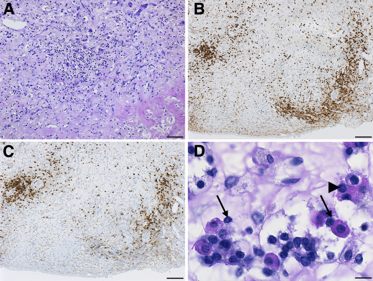
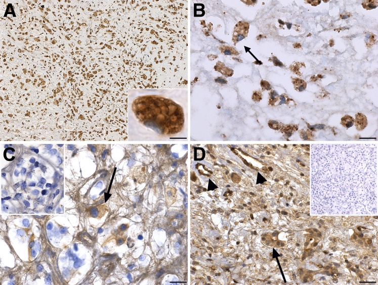
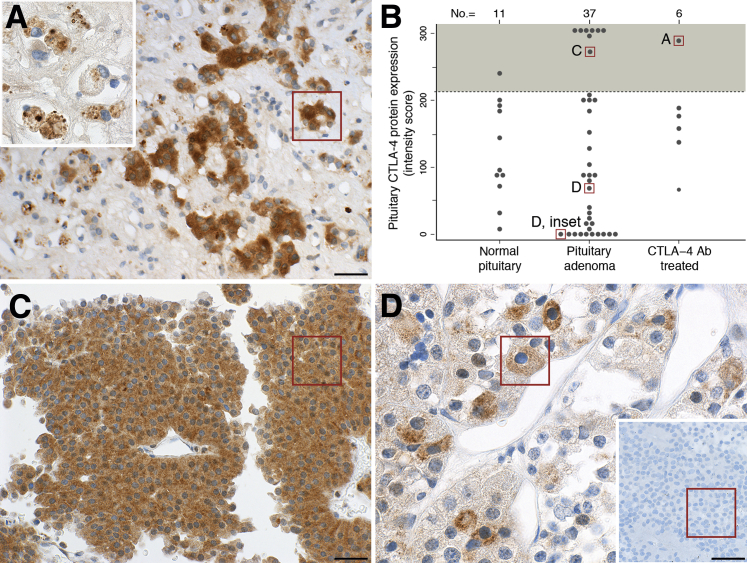
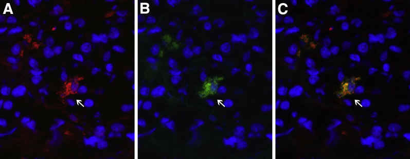
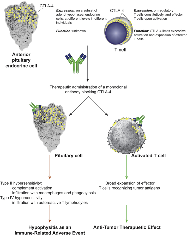
Hypophysitis that develops in cancer patients treated with monoclonal antibodies blocking cytotoxic T-lymphocyte-associated protein 4 (CTLA-4; an inhibitory molecule classically expressed on T cells) is now reported at an incidence of approximately 10%. Its pathogenesis is unknown, in part because no pathologic examination of the pituitary gland has been reported to date. We analyzed at autopsy the pituitary glands of six cancer patients treated with CTLA-4 blockade, one with clinical and pathologic evidence of hypophysitis, one with mild lymphocytic infiltration in the pituitary gland but no clinical signs of hypophysitis, and four with normal pituitary structure and function. CTLA-4 antigen was expressed by pituitary endocrine cells in all patients but at different levels. The highest levels were found in the patient who had clinical and pathologic evidence of severe hypophysitis. This high pituitary CTLA-4 expression was associated with T-cell infiltration and IgG-dependent complement fixation and phagocytosis, immune reactions that induced an extensive destruction of the adenohypophyseal architecture. Pituitary CTLA-4 expression was confirmed in a validation group of 37 surgical pituitary adenomas and 11 normal pituitary glands. The study suggests that administration of CTLA-4 blocking antibodies to patients who express high levels of CTLA-4 antigen in the pituitary can cause an aggressive (necrotizing) form of hypophysitis through type IV (T-cell dependent) and type II (IgG dependent) immune mechanisms.


Figures

Supplemental Figure S1
Cranial magnetic resonance imaging appearance of the index case at diagnosis. A: T2-weighted image showing a slightly enlarged pituitary gland (6.1 mm in height). B: T1-weighted image after contrast showing the presence of large areas of low intensity (arrows) inside the anterior pituitary, consistent with necrosis.
Supplemental Figure S2
Histopathology of autopsy case 6. A: Hematoxylin and eosin staining shows the mild lymphocytic infiltration (arrows) into the pituitary acini, with initial damage to the endocrine cells (inset). B: Immunohistochemical staining shows the presence of scattered CD3-positive T cells (arrows) into the pituitary parenchyma. The inset shows the typical cytoplasmic appearance of a T cell stained for CD3. Original magnification: ×20 (A and B, main images); ×40 (A and B, insets).
Supplemental Figure S3
Immunohistochemical staining for Foxp3. A: Pituitary of the index autopsy case. The inset shows that Foxp3 is not expressed in the index autopsy case. B: Pituitary from a melanoma patient who had received treatment with ipilimumab but had not developed hypophysitis. The inset shows that pituitary acini are negative for Foxp3. Boxed areas are enlarged in the insets. Original magnification: ×40 (A and B, main images); ×160 (A and B, insets).
Figure 1
Figure 1
Necrotizing hypophysitis induced by administration of a CTLA-4 blocking antibody. A: Hematoxylin and eosin (H&E) low power view of the anterior pituitary of the autopsy index case, to show a large necrotic area (red outline), the mononuclear cell infiltration (in the center of the field), and the rarity of endocrine acini remaining (black outline). B: Masson's trichrome staining to show (turquoise) the extensive fibrosis. C and D: H&E mid (C) and high (D) power view of one of the remaining areas containing pituitary endocrine cells, showing the marked infiltration with hematopoietic mononuclear cells (E) H&E mid power to show an island of remaining acidophils, that stain positive for growth hormone (F). Scale bars: 200 μm (A and B); 100 μm (C); 50 μm (D–F). Original magnification: ×20 (A and B); ×40 (C); ×100 (D); ×64 (E and F).
Figure 2
Figure 2
Evidence of type IV hypersensitivity mechanisms in the pituitary gland of the autopsy case. A: Diffuse infiltration of the anterior pituitary with lymphocytes. Lymphocytes are CD4-positive T cells (B) and CD20-positive B cells (C). D: Lymphocytes intimately contact the pituitary acidophil cells (arrows), and in some cases penetrate in their cytosol (arrowhead). Scale bars: 50 μm (A); 100 μm (B and C); 20 μm (D). Original magnification: ×64 (A); ×40 (B and C); ×260 (D).
Figure 3
Figure 3
Evidence of type II hypersensitivity mechanisms in the pituitary gland of the autopsy case. A: CD68 staining showing the marked infiltration of the gland with macrophages. The inset shows at high power a single macrophage with prominent vacuolated cytoplasm. B: High power view of prolactin staining to show that the positivity is found almost exclusively inside the cytosol of macrophages. C: Binding of IgG2 immunoglobulins to pituitary endocrine cells (arrow) of the case. The inset shows at the same magnification the absence of staining for IgG1. D: Deposition of complement C4d on some pituitary endocrine cells (arrow) and endothelial cells (arrowheads) of the case. The inset shows the absence of Cd4 deposition in a control pituitary. Scale bars: 100 μm (A); 20 μm (B and C); 50 μm (D). Original magnification: ×40 (A, main image); ×260 (A, inset, B, and C, main image and inset); ×160 (D, main image and inset).
Figure 4
Figure 4
Expression of pituitary CTLA-4 in the human pituitary gland at autopsy. A: In the index case, the few remaining acini of endocrine cells in the pituitary strongly express CTLA-4. The inset shows that CTLA-4–positive granules are found inside the cytosol of infiltrating macrophages. B: Histopathologic score to quantify the expression of pituitary CTLA-4 protein in the study population; lettered boxes correlate to the boxed areas in the indicated panels. The shaded area above the dotted lines indicates the specimens that express CTLA-4 at high intensity in most endocrine pituitary cells. C: A pituitary adenoma expressing CTLA-4 strongly. D: A pituitary adenoma expressing CTLA-4 at moderate levels in a subset of endocrine cells; the inset shows a pituitary adenoma negative for CTLA-4. Scale bars: 50 μm (A and C); 20 μm (D). Original magnifications: ×100 (A, main image); ×260 (A, inset, and D, main image); ×160 (C and D, inset).
Figure 5
Figure 5
Double indirect immunofluorescence using the pituitary of autopsy case 6 as substrate and antibodies to follicle-stimulating hormone (FSH)-β and CTLA-4. A: Staining with anti–FSH-β identifies a clear positive cell (arrows). B: The same cell is also stained with anti–CTLA-4. C: Merging of the red (FSH-β) and green (CTLA-4) channel yields a yellowish cell indicating colocalization. Original magnificaiton, ×40 (A–C).
Figure 6
Figure 6
Schematic representation of the mechanism of action of CTLA-4 blocking antibodies. Administration of a CTLA-4 blocking antibody binds to CTLA-4 expressed on T lymphocytes, leading to the beneficial expansion of effector T cells that recognize novel tumor antigens and eliminate the tumor (right side). The same administered antibody, however, also binds to CTLA-4 expressed on pituitary cells, leading to type II and type IV hypersensitivity mechanisms that cause hypophysitis and pituitary cytotoxicity (left side).

References

    1. Leporati P., Landek-Salgado M.A., Lupi I., Chiovato L., Caturegli P. IgG4-related hypophysitis: a new addition to the hypophysitis spectrum. J Clin Endocrinol Metab. 2011;96:1971–1980. - PMC - PubMed
    1. Caturegli P., Iwama S. From Japan with love: another tessera in the hypophysitis mosaic. J Clin Endocrinol Metab. 2013;98:1865–1868. - PubMed
    1. Caturegli P., Newschaffer C., Olivi A., Pomper M.G., Burger P.C., Rose N.R. Autoimmune hypophysitis. Endocr Rev. 2005;26:599–614. - PubMed
    1. Deodhare S.S., Bilbao J.M., Kovacs K., Horvath E., Nomikos P., Buchfelder M., Reschke K., Lehnert H. Xanthomatous hypophysitis: a novel entity of obscure etiology. Endocr Pathol. 1999;10:237–241. - PubMed
    1. van der Vliet H.J., Perenboom R.M. Multiple pseudotumors in IgG4-associated multifocal systemic fibrosis. Ann Intern Med. 2004;141:896–897. - PubMed

Publication types

MeSH terms