Review
. 2016 Oct 18;20(1):279.
doi: 10.1186/s13054-016-1458-3.

The digestive tract as the origin of systemic inflammation

Affiliations
Review

The digestive tract as the origin of systemic inflammation

Petrus R de Jong et al. Crit Care. .

Abstract

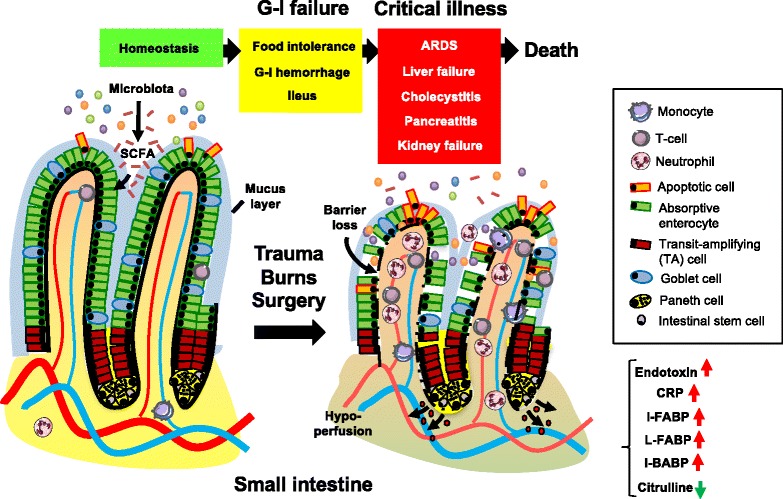
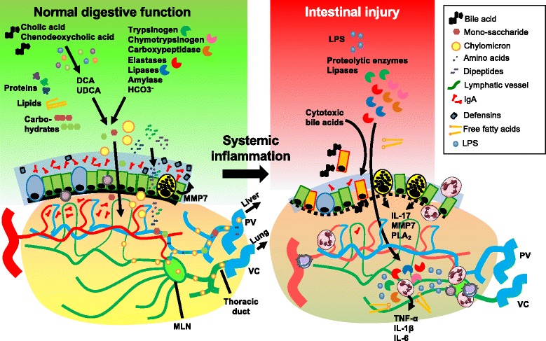
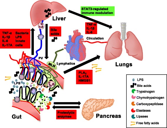
Failure of gut homeostasis is an important factor in the pathogenesis and progression of systemic inflammation, which can culminate in multiple organ failure and fatality. Pathogenic events in critically ill patients include mesenteric hypoperfusion, dysregulation of gut motility, and failure of the gut barrier with resultant translocation of luminal substrates. This is followed by the exacerbation of local and systemic immune responses. All these events can contribute to pathogenic crosstalk between the gut, circulating cells, and other organs like the liver, pancreas, and lungs. Here we review recent insights into the identity of the cellular and biochemical players from the gut that have key roles in the pathogenic turn of events in these organ systems that derange the systemic inflammatory homeostasis. In particular, we discuss the dangers from within the gastrointestinal tract, including metabolic products from the liver (bile acids), digestive enzymes produced by the pancreas, and inflammatory components of the mesenteric lymph.

Keywords: Acute inflammation; Gastrointestinal failure; Gut-liver crosstalk; Pancreatitis.


Figures

Fig. 1
Fig. 1
Pathogenic events in the course of gastro-intestinal failure. Potential toxic components in the small intestine are contained in the lumen under homeostatic conditions, whilst enterocytes allow the uptake of nutrients such as short-chain fatty acids (SCFA) across the epithelial barrier. In the critically ill patient suffering from major trauma or burns or undergoing surgery, protective mechanisms that maintain the gut barrier fail due to circulatory and neuroendocrine dysregulation. This results in gastro-intestinal (G-I) failure, which coincides with clinical signs such as oral intolerance, gastrointestinal hemorrhage, or ileus. G-I failure is a progressive clinical syndrome in which an early stage of predominantly gastrointestinal symptoms may be followed by extraintestinal derangements, such as acute respiratory distress syndrome (ARDS), liver failure, cholecystitis, pancreatitis, or kidney failure. G-I failure is associated with epithelial denudation, villus shortening, and inflammatory cell infiltration in the small intestine at the tissue level. Finally, various clinical biomarkers may aid in the diagnosis of G-I failure, including plasma or urinary levels of intestinal-type fatty acid-binding protein (I-FABP), liver-type fatty acid-binding protein (L-FABP), ileal bile acid-binding protein (I-BABP) or citrulline. CRP C-reactive protein
Fig. 2
Fig. 2
Cellular and molecular players in intestinal injury caused by systemic inflammation. A large variety of pancreatic and hepatobiliary products are involved in the complex digestive function of the small intestine, e.g., proteolytic enzymes, lipases, amylases, bicarbonate, and primary bile acids. The primary bile acids (cholic acid, chenodeoxycholic acid) are converted to secondary bile acids (e.g., lithocholic acid, DCA, UDCA) by the microbiota. The digestive enzymes derived from the host or microbiota ensure the breakdown of macromolecules to soluble nutrients, which is followed by uptake and transport to the portal circulation or intestinal lymph tract. While the intestinal microflora are crucial for these digestive functions in the lumen, their quantity and topography are tightly controlled by the thick mucus layer that contains secretory IgA and antimicrobial peptides (e.g., defensins) produced by mucosal immune cells and the epithelium. In critical illness, circulatory derangement results in gut barrier loss and the translocation of digestive enzymes, cytotoxic bile acids, free fatty acids (FFAs), and microbial substrates to the submucosa, which exacerbates local and systemic inflammatory reactions. The intestinal epithelium is also an important source of pro-inflammatory mediators that are released into the circulation, including IL-17, lipid mediators produced by phospholipase A2 (PLA 2), and antimicrobial peptides derived from Paneth cells. These pathogenic events also pave the way for cytotoxic components to directly leak from the intestinal lumen to mesenteric lymph vessels, which constitutes the gut–lymph–lung axis. MLN mesenteric lymph node, MMP matrix metalloproteinase, PV portal vein, VC vena cava
Fig. 3
Fig. 3
Direct and indirect interactions between the gut and other organs in systemic inflammation. Luminal components of the small intestine can spread to the circulation via the portal vein (PV) and liver. This includes pro-inflammatory constituents such as lipopolysaccharide (LPS), bacterial DNA, whole bacteria, other bacterial products, and free fatty acids (FFAs). Toxic components of the gut lumen, including FFAs, inflammatory products of phospholipase A2 (PLA 2), pro-inflammatory cytokines (e.g., IL-17), and damage-associated molecular substrates such as high mobility group box 1 protein (HMGB1), can reach the pulmonary circulation via mesenteric lymph. Bile acids from the liver, including cholic acid, DCA, and chenodeoxycholic acid, mediate cytotoxic effects on intestinal epithelial cells. STAT3 signaling in Kupffer cells (KCs) of the liver maintains tolerance under homeostatic conditions, whereas KCs produce high levels of pro-inflammatory cytokines in systemic inflammation with toxic effects on the lung parenchyma. Finally, activated serine proteases, elastases, and lipases produced by the pancreas can cause local tissue destruction and activation of immune cells in intestinal tissues, leading to exacerbated systemic inflammatory responses

References

    1. van der Flier LG, Clevers H. Stem cells, self-renewal, and differentiation in the intestinal epithelium. Annu Rev Physiol. 2009;71:241–60. doi: 10.1146/annurev.physiol.010908.163145. - DOI - PubMed
    1. Sato T, van Es JH, Snippert HJ, Stange DE, Vries RG, van den Born M, Barker N, Shroyer NF, van de Wetering M, Clevers H. Paneth cells constitute the niche for Lgr5 stem cells in intestinal crypts. Nature. 2011;469(7330):415–8. doi: 10.1038/nature09637. - DOI - PMC - PubMed
    1. Clevers H, Batlle E. SnapShot: the intestinal crypt. Cell. 2013;152(5):1198. doi: 10.1016/j.cell.2013.02.030. - DOI - PubMed
    1. Vaishnava S, Behrendt CL, Ismail AS, Eckmann L, Hooper LV. Paneth cells directly sense gut commensals and maintain homeostasis at the intestinal host-microbial interface. Proc Natl Acad Sci U S A. 2008;105(52):20858–63. doi: 10.1073/pnas.0808723105. - DOI - PMC - PubMed
    1. Tremaroli V, Backhed F. Functional interactions between the gut microbiota and host metabolism. Nature. 2012;489(7415):242–9. doi: 10.1038/nature11552. - DOI - PubMed

MeSH terms

Substances

LinkOut - more resources