

. 2016 Sep;68 Suppl 2(Suppl 2):S170-S174.
doi: 10.1016/j.ihj.2015.07.043.
Epub 2016 Jan 18.
Late presentation of an unruptured giant sub-mitral lateral wall true left ventricular aneurysm
Affiliations
- PMID: 27751279
- PMCID: PMC5067458
- DOI: 10.1016/j.ihj.2015.07.043
Item in Clipboard
Late presentation of an unruptured giant sub-mitral lateral wall true left ventricular aneurysm
Indian Heart J.
2016 Sep.
No abstract available
Keywords: Lateral wall; Left ventricular aneurysm; Unruptured.
Figures

Lateral wall MI (diagnosed 2 years prior to aneurysm).

ECG showing sinus tachycardia and LVH strain pattern.

TTE of the patient showing LA, LV and the blood flow across the aneurysm in Parasternal Long axis view.

TTE on shallow four-chamber view showing the size of the aneurysm along with other structures LV, RA, and RV.

TTE showing blood flow across the neck of the aneurysm.

TTE shallow two chamber view showing LA, LV, and aneurysm.

PLAX view of TTE showing thinning of LV.

TTE showing the LA, LV and the measurements of the aneurysm having dimensions 6.84 and 6.43 cm and having an area of 35.5 cm2.
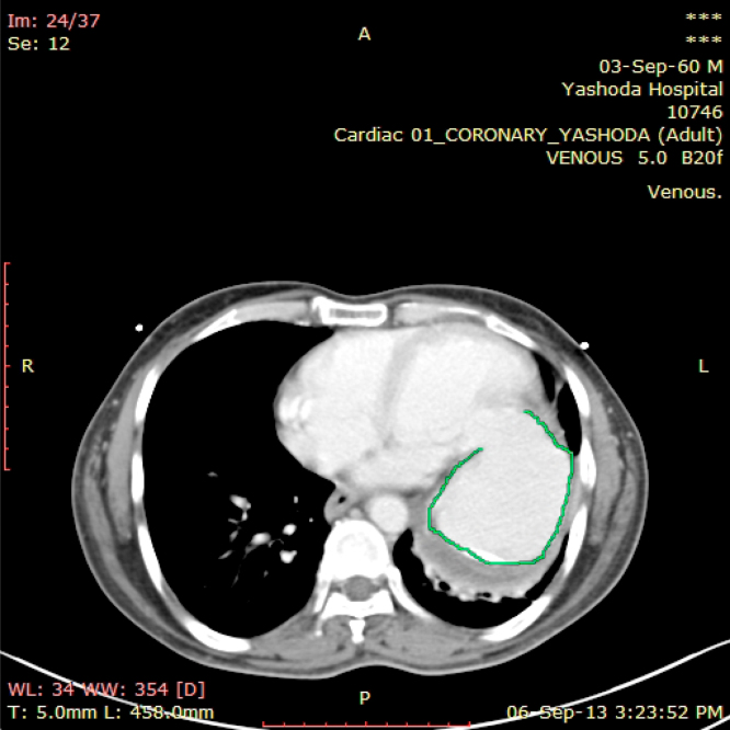
CT Angiogram marking the borders of the aneurysm.

CTA showing the dimensions of the aneurysms in axial plane.

CTA showing the dimensions of the aneurysms in coronal plane.

VRT images on CT angiogram showing big aneurysm attached to LV.

VRT images on CT angiogram showing big aneurysm attached to LV.

VRT images on CT angiogram showing big aneurysm attached to LV.
Similar articles
-
Giant ischemic left ventricular submitral aneurysm.Indian Heart J. 2016 Sep;68 Suppl 2(Suppl 2):S168-S169. doi: 10.1016/j.ihj.2015.10.383. Epub 2016 Jan 13. Indian Heart J. 2016. PMID: 27751278 Free PMC article. No abstract available.
-
Giant left ventricular outflow tract aneurysm presenting as unstable angina: an unusual case.J Am Coll Cardiol. 2014 Apr 8;63(13):1332. doi: 10.1016/j.jacc.2013.09.086. Epub 2014 Feb 13. J Am Coll Cardiol. 2014. PMID: 24503124 No abstract available.
-
[Off-pump left ventricular reconstruction for giant aneurysm in patient with low myocardial capacity].Khirurgiia (Mosk). 2017;(9):85-87. doi: 10.17116/hirurgia2017985-87. Khirurgiia (Mosk). 2017. PMID: 28914839 Russian. No abstract available.
-
Endoaneurysmorrhaphy for a Giant Inferobasal Left Ventricular Aneurysm Restoring Mitral Function.J Heart Valve Dis. 2017 Sep;26(5):613-615. J Heart Valve Dis. 2017. PMID: 29762937
-
Giant aneurysm arising from the left atrial branch of the left circumflex artery and rupturing into the right atrium.Eur Heart J Cardiovasc Imaging. 2013 Jun;14(6):549. doi: 10.1093/ehjci/jes286. Epub 2012 Dec 18. Eur Heart J Cardiovasc Imaging. 2013. PMID: 23250890 No abstract available.
References
-
- Dolly C.H., Dotter C.T., Steinberg H. Ventricular aneurysm in a 29-year-old man studied angiocardiographically. Am Heart J. 1951;42:894–899. - PubMed
-
- Bohdiewicz P.J. Single photon emission computed tomography radionuclide ventriculography in the noninvasive diagnosis and evaluation of a false left ventricular aneurysm (Pseudonaeurysm) Clin Nucl Med. 2003;28:821–826. - PubMed
-
- Brown S.L., Gropler R.J., Harris K.M. Distinguishing left ventricular aneurysm from pseudoaneurysm: a review of the literature. CHEST. 1997;111:1403–1409. - PubMed
-
- Frances C., Romero A., Grady D. Left ventricular pseudoaneurysm. J Am Coll Cardiol. 1998;32:557–561. - PubMed
MeSH terms
LinkOut - more resources
Full Text Sources
Other Literature Sources
Medical
