

Successful management of a giant unruptured mycotic coronary artery aneurysm after coronary angioplasty
- PMID: 27751325
- PMCID: PMC5067809
- DOI: 10.1016/j.ihj.2016.08.006
Successful management of a giant unruptured mycotic coronary artery aneurysm after coronary angioplasty
Abstract
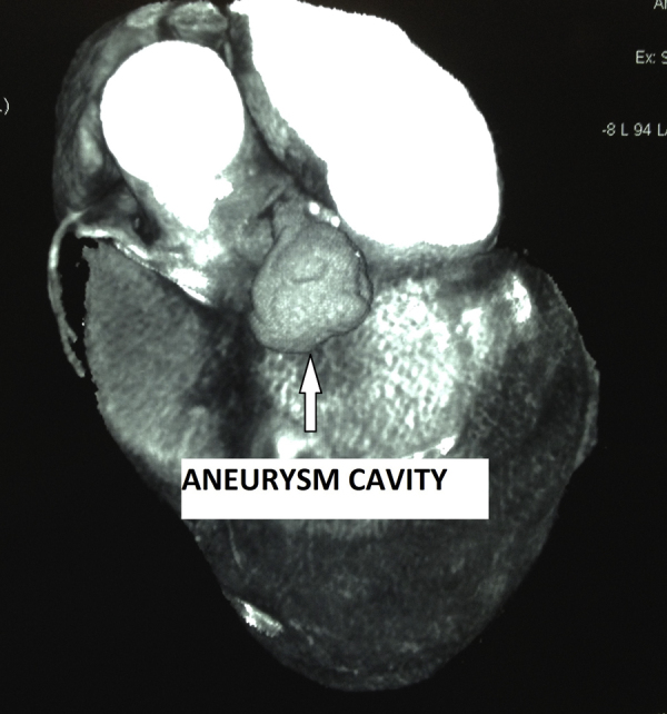
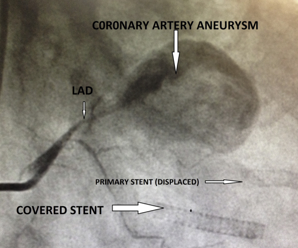
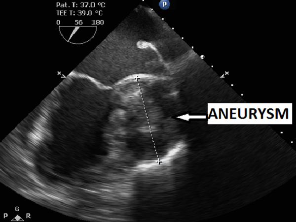
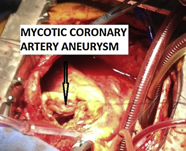
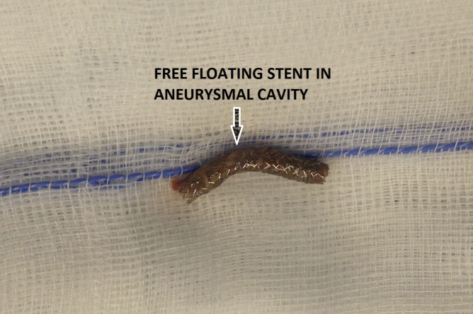
Coronary artery stent infection has been reported with both bare metal stent and drug eluting stent and can present as mycotic coronary artery aneurysm, pseudoaneurysm, myocardial abscess, pericarditis or exudative effusion. Infection at the site of coronary stent implantation is rare and is believed to result typically from either direct stent contamination at the time of delivery or transient bacteraemia from access site. Introduction of drug-eluting stent (DES) has led to a marked reduction in the problem of in-stent restenosis across all patient subsets and lesions complexities. Recently, several case reports of pseudoaneurysm formation after DES implantation have been reported in the literature. We describe the successful surgical management of giant mycotic pseudoaneurysm of left anterior descending artery (LAD) presenting as fever of unknown origin. This report illustrates the importance of early detection and prompt management of these rare coronary pseudoaneurysms, which is a highly lethal condition.
Keywords: Mycotic coronary artery aneurysm.
Copyright © 2016 Cardiological Society of India. Published by Elsevier B.V. All rights reserved.
Figures
Similar articles
-
Infected pseudoaneurysm involving a drug-eluting stent.Interact Cardiovasc Thorac Surg. 2011 Apr;12(4):636-7. doi: 10.1510/icvts.2010.257337. Epub 2011 Jan 12. Interact Cardiovasc Thorac Surg. 2011. PMID: 21228044
-
Coronary artery aneurysms after drug-eluting stent implantation.Interact Cardiovasc Thorac Surg. 2011 Apr;12(4):638. doi: 10.1510/icvts.2010.257337B. Interact Cardiovasc Thorac Surg. 2011. PMID: 21429891 No abstract available.
-
Is the pathology different between drug-eluting stent and bare stent related coronary aneurysms?Interact Cardiovasc Thorac Surg. 2011 Apr;12(4):638. doi: 10.1510/icvts.2010.257337A. Interact Cardiovasc Thorac Surg. 2011. PMID: 21429892 No abstract available.
-
Coronary Artery Aneurysm Caused by a Stent Fracture.Int Heart J. 2018;59(1):203-208. doi: 10.1536/ihj.17-081. Int Heart J. 2018. PMID: 29375112 Review.
-
Fatal infection of coronary stent implantation.Cathet Cardiovasc Diagn. 1996 Oct;39(2):168-70; discussion 171. doi: 10.1002/(SICI)1097-0304(199610)39:2<168::AID-CCD12>3.0.CO;2-D. Cathet Cardiovasc Diagn. 1996. PMID: 8922319 Review.
Cited by
-
Commentary: Coronary artery mycotic aneurysm in a patient suffering from subacute endocarditis: a case report and literature review.Front Cardiovasc Med. 2023 Nov 28;10:1286416. doi: 10.3389/fcvm.2023.1286416. eCollection 2023. Front Cardiovasc Med. 2023. PMID: 38089769 Free PMC article. No abstract available.
-
Stenotrophomonas maltophilia pericarditis.Anatol J Cardiol. 2017 Dec;18(6):439-440. doi: 10.14744/AnatolJCardiol.2017.8024. Anatol J Cardiol. 2017. PMID: 29256885 Free PMC article. No abstract available.
-
Ruptured mycotic coronary artery aneurysm.BMJ Case Rep. 2023 Apr 12;16(4):e254488. doi: 10.1136/bcr-2022-254488. BMJ Case Rep. 2023. PMID: 37045550 Free PMC article.
-
Management of a Mycotic Right Coronary Artery Aneurysm With Contained Rupture.JACC Case Rep. 2022 Jun 1;4(11):694-698. doi: 10.1016/j.jaccas.2022.03.018. eCollection 2022 Jun 1. JACC Case Rep. 2022. PMID: 35677799 Free PMC article.
-
Mycotic and non-mycotic coronary artery aneurysms-A review of the rarity.J Clin Imaging Sci. 2022 Mar 30;12:13. doi: 10.25259/JCIS_218_2021. eCollection 2022. J Clin Imaging Sci. 2022. PMID: 35414960 Free PMC article.
References
-
- Mylonakis E., Calderwood S.B. Infective endocarditis in adults. N Engl J Med. 2001;345:1318–1330. - PubMed
-
- Osevala M.A., Heleotis T.L., Dejene B.A. Successful treatment of a ruptured mycotic coronary artery aneurysm. Ann Thorac Surg. 1999;67:1780–1782. - PubMed
-
- Briguori C., Sarais C., Sivieri G. Polytetrafluoroethylene-covered stent and coronary artery aneurysms. Catheter Cardiovasc Interv. 2002;55:326–330. - PubMed
-
- Chen I.C., Chao T.H., Wu Y.H., Kan C.D., Fang C.C. Afebrile mycotic aneurysm with rupture in right coronary artery after bare-metal stent implantation: a case report and review of literature. Acta Cardiol Sin. 2012;28:344–348.
-
- Matsumoto M., Konishi Y., Miwa S. Mycotic aneurysm of the left coronary artery. Ann Thorac Surg. 1998;65:841–842. - PubMed
Publication types
MeSH terms
LinkOut - more resources
Full Text Sources
Other Literature Sources
