. 2016 Oct;9(5):419-430.
doi: 10.1016/j.tranon.2016.07.008.

Effect of Repeat Dosing of Engineered Oncolytic Herpes Simplex Virus on Preclinical Models of Rhabdomyosarcoma

Affiliations

Effect of Repeat Dosing of Engineered Oncolytic Herpes Simplex Virus on Preclinical Models of Rhabdomyosarcoma

Alicia M Waters et al. Transl Oncol. 2016 Oct.

Abstract

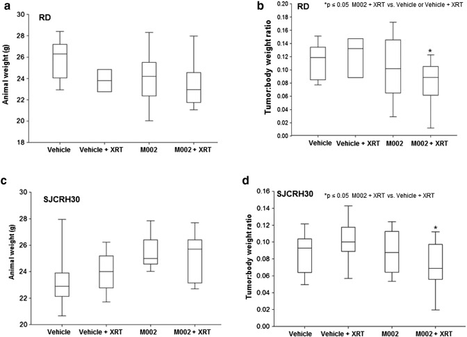
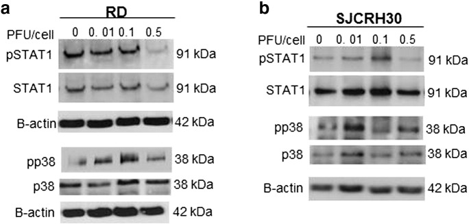
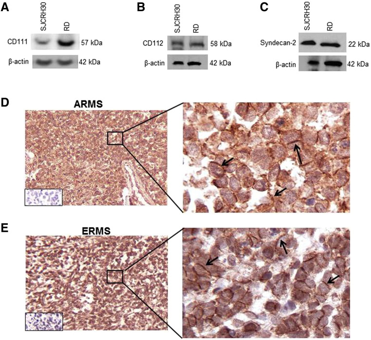
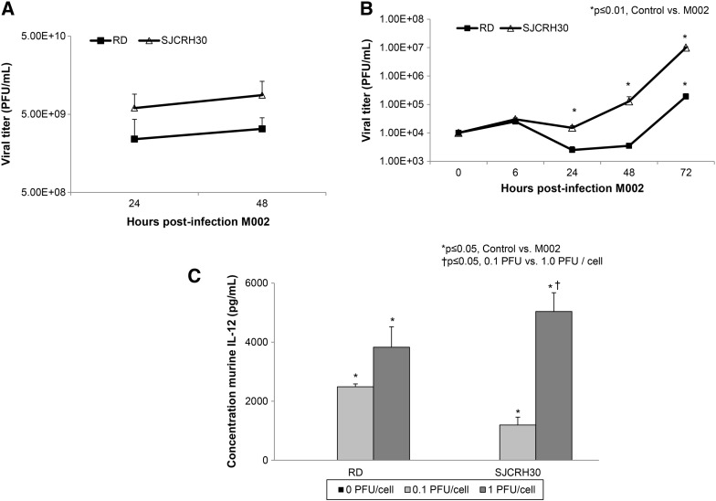
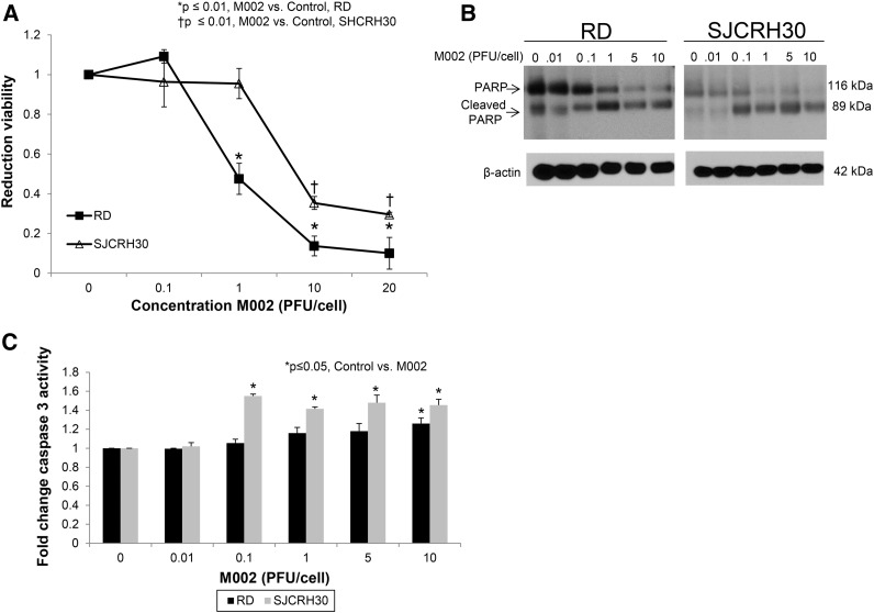
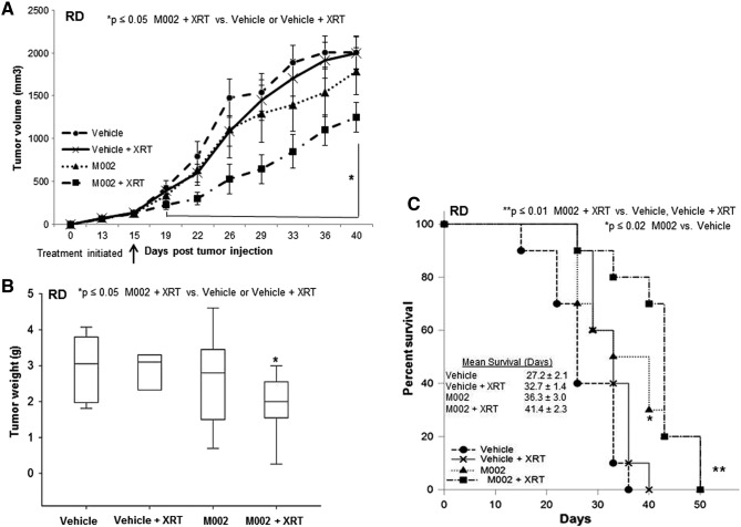
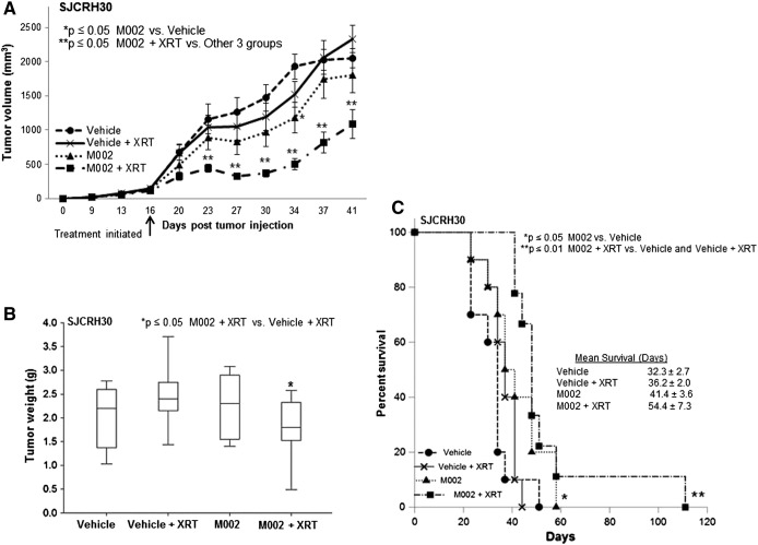
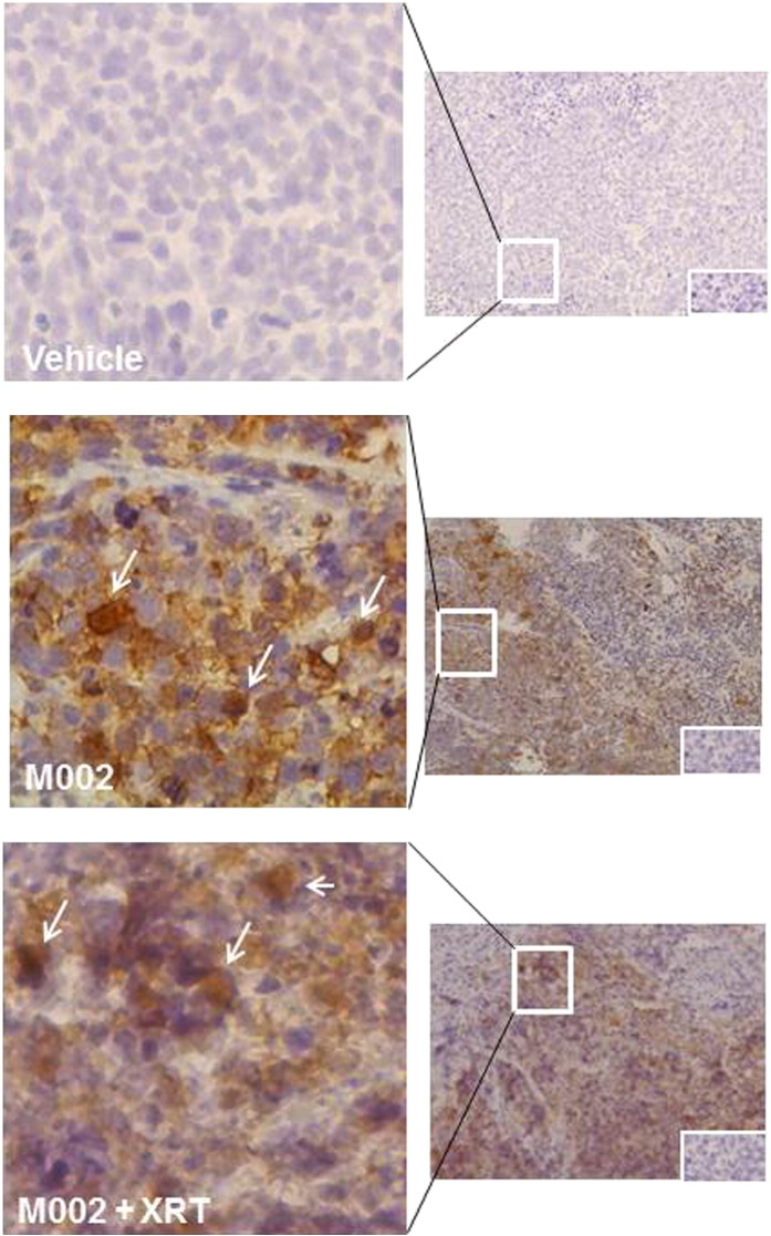
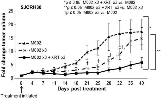
Rhabdomyosarcoma (RMS), a tumor of skeletal muscle origin, is the most common sarcoma of childhood. Despite multidrug chemotherapy regimens, surgical intervention, and radiation treatment, outcomes remain poor, especially in advanced disease, and novel therapies are needed for the treatment of these aggressive malignancies. Genetically engineered oncolytic viruses, such as herpes simplex virus-1 (HSV), are currently being explored as treatments for pediatric tumors. M002, an oncolytic HSV, has both copies of the γ134.5 gene deleted, enabling replication in tumor cells but thwarting infection of normal, postmitotic cells. We hypothesized that M002 would infect human RMS tumor cells and lead to decreased tumor cell survival in vitro and impede tumor growth in vivo. In the current study, we demonstrated that M002 could infect, replicate in, and decrease cell survival in both embryonal (ERMS) and alveolar rhabdomyosarcoma (ARMS) cells. Additionally, M002 reduced xenograft tumor growth and increased animal survival in both ARMS and ERMS. Most importantly, we showed for the first time that repeated dosing of oncolytic virus coupled with low-dose radiation provided improved tumor response in RMS. These findings provide support for the clinical investigation of oncolytic HSV in pediatric RMS.


Figures

Figure S1
Figure S1
Animal weights at euthanasia in RD (A) and SJCRH30 (C) human RMS xenografts. The median weights of the animals were not uniform at the time of euthanasia. Lines in the boxes represent median measurements, and whiskers represent the 5th and 95th percentile. A calculation of tumor:body weight ratio was completed for the RD (B) and the SJCRH30 (D) xenografts to determine if animal weight affected the tumor size. The tumor:body weight ratios correlated positively with the data of the tumor weights alone, effectively eliminating animal size as a confounding factor on tumor growth.
Figure S2
Figure S2
Phosphorylation of p38 and STAT1. (A) RD and (B) SJCRH30 cells were treated with increasing MOI of M002, and immunoblotting for total and phosphorylated Stat1 and p38 was performed. Phosphorylation of Stat1 was not increased following M002 treatment in either cell line. Phosphorylation of p38 was increased in both cell lines following M002 treatment. Immunoblotting for β-actin was completed as a loading control.
Figure 1
Figure 1
HSV entry receptors in cell lines and human RMS specimens. (A–C) Immunoblotting with CD111, CD112, and syndecan-2 specific antibodies demonstrated all three proteins to be present in whole cell lysates of SJCRH30 and RD human RMS cell lines. β-Actin was used to confirm equal protein loading. Immunohistochemical staining was performed on human ARMS (D) and ERMS (E) specimens. Representative photomicrographs at 10× and 40× are presented. There was CD111 staining present on the cell surface (black arrows) in both histologic types. Negative controls reacted appropriately (bottom left inserts, D, E).
Figure 2
Figure 2
Infectivity of M002 in RMS cell lines. (A) Single-step in vitro replication of M002. Monolayers of RD and SJCRH30 cells were infected with M002 at an MOI of 10 PFU/cell. Replicate cultures were harvested at 24 and 48 hours postinfection, and virus titers were determined on Vero cell monolayers. Mean virion yields were determined in four replicates at each time point, and standard error of the mean was determined. By 24 hours postinfection, there were significant viral titers noted in both cell lines that continued to increase at 48 hours postinfection. (B) Multistep replication of M002. Monolayers of RD and SJCRH30 cells were infected with M002 at an MOI of 0.1 PFU/cell, and at 6, 24, 48, and 72 hours postinfection, supernates were collected, and virus titers were determined on Vero cell monolayers. Mean virion yields were determined in four replicates at each time point, and standard error of the mean was determined. In the RD cell line, M002 replicated more than a log higher than control at 72 hours postinfection, and in the SJCRH30 cell line, replication of virus was more than 3 logs greater than time zero. (C) Because M002 was engineered to produce mIL-12, to further verify infection, mIL-12 production was determined in RD and SJCRH30 cell lines following treatment with M002 oHSV. Cell lines were infected with M002 at 0, 0.1, or 1.0 PFU/cell. At 48 hours postinfection, the supernates were collected, and concentrations of mIL-12 were determined by ELISA. Data are reported as mean ± standard error of the mean. There was a significant increase in mIL-12 production in both cell lines even with the lower MOI of virus.
Figure 3
Figure 3
Treatment with M002 resulted in cell death. (A) RD and SJCRH30 cell lines were treated with M002 at increasing MOI. After 72 hours of treatment, cell viability was measured with alamarBlue assays. Data are reported as mean ± standard error of the mean. There was a significant decrease in viability in both cell lines following M002 treatment. The LD50 was calculated for each cell line for M002 and was 3.3 ± 0.1 PFU/cell for embryonal RD cells and 4.1 ± 0.4 PFU/cell for the alveolar SJCRH30 cells. (B) To determine whether RMS cells were undergoing an apoptotic process following M002 treatment, immunoblotting for cleavage of PARP was completed. There were a significant decrease in total PARP staining and an increase in cleaved PARP staining in both cell lines with increasing MOIs. (C) To further verify apoptosis, a caspase 3 activation kit was utilized. There was a significant increase in caspase 3 activation following M002 treatment, indicating that the RMS cells were undergoing an apoptotic process.
Figure 4
Figure 4
M002 treatment of RD xenografts. (A) RD human RMS tumor cells (2.5 × 106 cells) were injected into the right flank of athymic nude mice. Once tumors reached a volume of 250 mm3, animals received an intratumoral injection of vehicle (PBS+ 10% glycerol, 50 μl [n = 20]) or M002 virus (1 × 107 PFU/50 μl [n = 20]). Half of the animals in each group also received low-dose irradiation (3 Gy) at the time of injection. Tumor volumes were measured twice weekly [(width)2 × length]/2 for 40 days. Data are reported as tumor volume ± standard error mean. In the animals treated with M002 and XRT, there was a significant decrease in tumor growth beginning at day 19 when compared with the animals in the vehicle or vehicle + XRT treatment groups. Tumor volumes in the M002 + XRT were also smaller than those from M002 alone, but this did not reach statistical significance. The addition of low-dose (3 Gy) XRT to the vehicle treatment did not significantly affect tumor growth (Figure 3A). (B) Xenograft tumors were weighed at euthanasia. There was a significant decrease in tumor weight in the animals treated with M002 and XRT when compared with animals that received vehicle alone or vehicle with XRT. Tumor weights in M002 + XRT group tended to be less than those of M002 alone but did not reach statistical significance. Lines in bars represent the median, and the whiskers represent the 5th and 95th percentile. (D) Kaplan-Meier curves were constructed with log-rank statistics to evaluate animal survival. Animals treated with M002 had significantly increased survival over those treated with vehicle alone. Animals treated with M002 + XRT also had a significant survival advantage when compared with those treated with vehicle or vehicle +XRT.
Figure 5
Figure 5
M002 treatment of SJCRH30 xenografts. (A) SJCRH30 cells (2.0 × 106 cells) were injected into the subcutaneous space of the right flank of female nude mice (n = 40). Once tumors reached 250 mm3, animals received an intratumoral injection of vehicle (PBS+ 10% glycerol, 50 μl [n = 20]) or M002 virus (1 × 107 PFU/50 μl [n = 20]). Half of each group was also treated with 3-Gy external beam radiation at the time of injection (XRT). Tumor volumes were measured twice weekly and reported as tumor volume ± standard error. Beginning on day 23, there was a significant decrease in tumor volume in the animals treated with combined M002 + XRT. (B) Xenograft tumors were weighed at euthanasia. There was a significant decrease in tumor weight in the animals treated with M002 and XRT when compared with animals that received vehicle with XRT. Lines in bars represent the median, and the whiskers represent the 5th and 95th percentile. (C) Kaplan-Meier curves were constructed with log-rank statistics to examine animal survival. Animals treated with M002 had significantly increased survival over those treated with vehicle alone. Animals treated with M002 + XRT also had a significant survival advantage when compared with those treated with vehicle or vehicle and XRT.
Figure 6
Figure 6
Histological examinations of RMS xenografts for HSV. The presence of HSV in the tumors following treatment was verified with immunohistochemical staining for HSV-1. Representative photomicrographs are at 20× (right) and 40× (left). Immunohistochemical staining for HSV-1 detected virus in tumors injected with M002 (middle panels brown stain, white arrows) and M002 + XRT (bottom panels brown stain, white arrows), present at 7 days post virus injection. Tumors injected with vehicle did not demonstrate HSV staining (upper panels). Negative controls reacted appropriately (insert, bottom right, upper, middle, and lower panels).
Figure 7
Figure 7
Repeated injections of M002 in SJCRH30 xenografts. SJCRH30 cells (2.0 × 106 cells) were injected into the subcutaneous space of the right flank of female nude mice (n = 20). Once tumors reached 250 mm3, animals were randomized to three groups: 1) single intratumoral injection of M002 virus (1 × 107 PFU/50 μl [n = 6]); 2) repeated intratumoral injection of M002 (1 × 107 PFU/50 μl) on days 0, 3, and 6 (n = 7); and 3) repeated intratumoral injection of M002 (1 × 107 PFU/50 μl) followed by low-dose irradiation (3 Gy, XRT) on days 0, 3, 6 and (n = 7). Tumor volumes were measured twice weekly and reported as fold change in tumor volume ± standard error. Beginning on posttreatment day 18, the animals receiving multiple doses of M002 had significantly less tumor growth than those treated with a single dose. The animals treated with multiple doses of M002 and XRT had significantly less tumor growth than either the single M002 dose or those that received multiple doses of M002 without XRT.

Similar articles

Cited by

References

    1. Rodeberg D, Paidas C. Childhood rhabdomyosarcoma. Semin Pediatr Surg. 2006;15:57–62. - PubMed
    1. Crist WM, Garnsey L, Beltangady MS, Gehan E, Ruymann F, Webber B, Hays DM, Wharam M, Maurer HM. Prognosis in children with rhabdomyosarcoma: a report of the Intergroup Rhabdomyosarcoma Studies I and II. Intergroup Rhabdomyosarcoma Committee. J Clin Oncol. 1990;8:443–452. - PubMed
    1. Rudzinski ER, Anderson JR, Hawkins DS, Skapek SX, Parham DM, Teot LA. The World Health Organization classification of skeletal muscle tumors in pediatric rhabdomyosarcoma: a report from the Children's Oncology Group. Arch Pathol Lab Med. 2015;139:1281–1287. - PMC - PubMed
    1. HaDuong JH, Martin AA, Skapek SX, Mascarenhas L. Sarcomas. Pediatr Clin North Am. 2015;62:179–200. - PubMed
    1. Arndt CA, Crist WM. Common musculoskeletal tumors of childhood and adolescence. N Engl J Med. 1999;341:342–352. - PubMed