. 2016 Dec:77:47-52.
doi: 10.1016/j.mcn.2016.10.004. Epub 2016 Oct 14.

Removal of aquaporin-4 from glial and ependymal membranes causes brain water accumulation

Affiliations

Removal of aquaporin-4 from glial and ependymal membranes causes brain water accumulation

Gry Fluge Vindedal et al. Mol Cell Neurosci. 2016 Dec.

Abstract

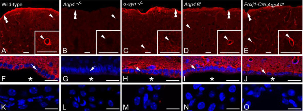
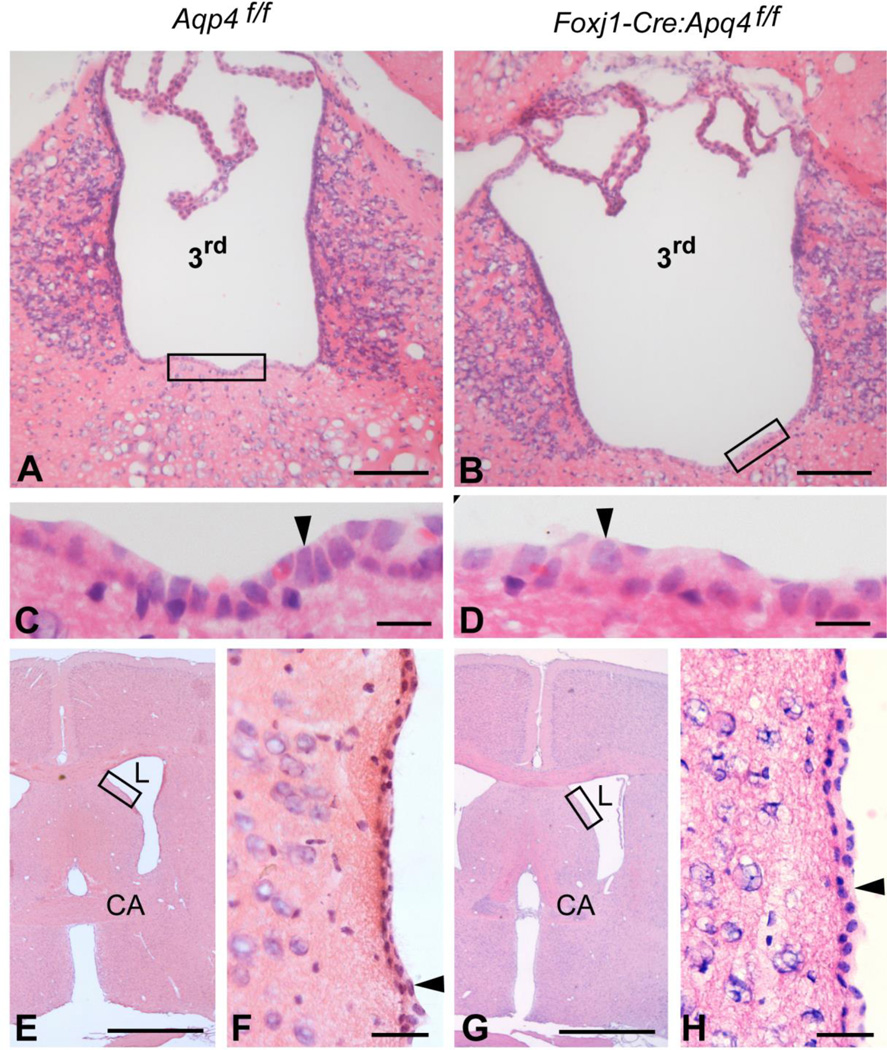
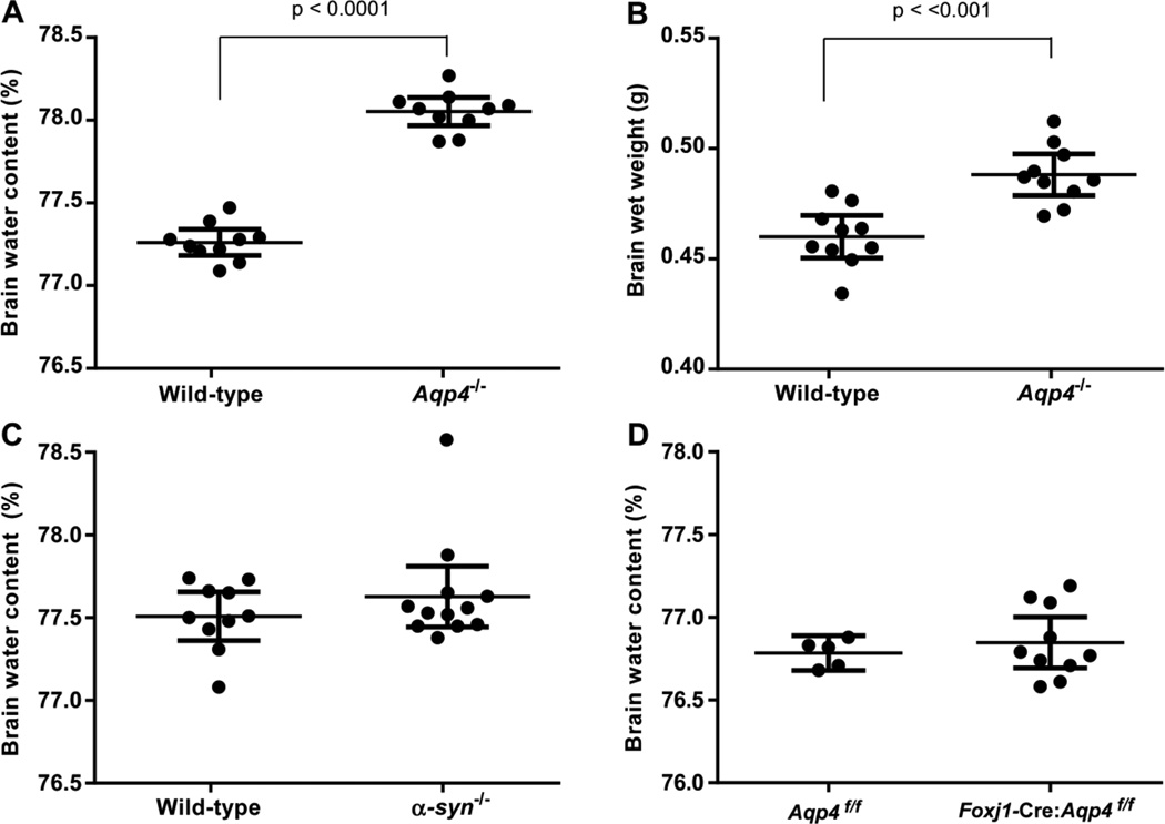
There is a constitutive production of water in brain. The efflux routes of this excess water remain to be identified. We used basal brain water content as a proxy for the capacity of water exit routes. Basal brain water content was increased in mice with a complete loss of aquaporin-4 (AQP4) water channels (global Aqp4-/- mice), but not in mice with a selective removal of perivascular AQP4 or in a novel mouse line with a selective deletion of ependymal AQP4 (Foxj1-Cre:Aqp4flox/flox mice). Unique for the global Aqp4-/- mice is the loss of the AQP4 pool subjacent to the pial membrane. Our data suggest that water accumulates in brain when subpial AQP4 is missing, pointing to a critical role of this pool of water channels in brain water exit.

Keywords: AQP4; Alpha-syntrophin; Aquaporin; Astrocytes; Brain edema; CSF; Cerebrospinal fluid; Endfeet; Ependyma; Extracellular space; Foxj1; Glia; Glymphatic; Interstitial fluid; Neuron-glial; Paravascular; Water homeostasis.


Figures

Figure 1
Figure 1
Effect of gene knockout on AQP4 distribution. AQP4 immunofluorence labeling (red) of the cortex (A-E), periventricular area (F-J; nuclear labeling in blue) and choroid plexus (K-O; nuclear labeling in blue) of wild-type (A, F, K), global Aqp4−/− mice (B, G, L), α-syntrophin−/− mice (C, H, M), homozygous Aqp4 floxed mice (“Aqp4f/f”) (D, I, N) and ependymal-conditional Aqp4−/− mice (”Foxj1-Cre:Aqp4f/f mice”) (E, J, O). A, F, K: In wild-type mice AQP4 labeling is most pronounced around cortical blood vessels (arrowhead; same vessel shown at higher magnification in inset) and along the pial surface (double arrowhead). Distinct AQP4 immunolabeling is evident in the basolateral membrane (arrow) of the ependyma covering the third ventricle (asterisk). The choroid plexus epithelium is devoid of AQP4 immunolabeling. B, G, L: Global Aqp4 deletion abolishes AQP4 immunolabeling. Labels as in A, F. C, H, M: In α-syntrophin−/− mice AQP4 labeling is lost around cortical blood vessels but still prominent along the pial surface and in the basolateral ependymal membrane. Labels as in A, F. D, I, N: AQP4 labeling in Aqp4f/f mice is indiscriminately from that of wild-types. Labels as in A, F. E, J, O: In Foxj1-Cre:Aqp4f/f mice the AQP4 immunofluorescence signal is lost from ependymal membranes but otherwise similar to that of litter controls (Aqp4f/fl mice) and wild-types. Labels as in A,F. Scale bars: 20 µm.
Figure 2
Figure 2
Ependymal-conditional Aqp4−/− mice do not reveal apparent abnormalities in the ependymal architecture. A–H: Hematoxylin-eosin staining of coronal brain sections revealed comparable morphology of the ependyma (arrowheads) lining the third (3rd) and lateral (L) ventricles in control (“Aqp4f/f”; A, C, E and F) and ependymal-conditional Aqp4−/− (”Foxj1-Cre:Aqp4f/f”; B, D, G and H) mice. Boxed areas in A, B, E and G are enlarged in C, D, F and H, respectively. CA, commisura anterior. Scale bars: A, B: 100 µm; C, D: 10 µm; E, G: 1 mm; F, H: 30 µm.
Figure 3
Figure 3
Impact of gene deletion on basal brain water content and brain wet weight. A, B: In Aqp4−/− mice (n=10) basal brain water content and brain wet weight are significantly elevated compared to wild-types (n=10; p values are indicated, unpaired t test). C, D: Neither α-syntrophin−/− mice (n=12) nor Foxj1-Cre:Aqp4f/f mice (n=10) differed significantly from their controls (n=10 and n=5, respectively) in basal brain water content (p>0.05 for both comparisons, unpaired t test).
Figure 4
Figure 4
Effect of intracisternal infusion of aCSF on ICP. Infusion of aCSF at 2 µl/min for 5 min (infusion period indicated by bar) elevates ICP in both Aqp4−/− and wild-types. Neither peak ICP increase nor ICP decay differed between the genotypes (p>0.05, unpaired t test for both comparisons).

Similar articles

Cited by

References

    1. Adams ME, Kramarcy N, Krall SP, Rossi SG, Rotundo RL, Sealock R, Froehner SC. Absence of alpha-syntrophin leads to structurally aberrant neuromuscular synapses deficient in utrophin. J Cell Biol. 2000;150:1385–1398. - PMC - PubMed
    1. Amiry-Moghaddam M, Otsuka T, Hurn PD, Traystman RJ, Haug FM, Froehner SC, Adams ME, Neely JD, Agre P, Ottersen OP, Bhardwaj A. An alpha-syntrophin-dependent pool of AQP4 in astroglial end-feet confers bidirectional water flow between blood and brain. Proc Natl Acad Sci U S A. 2003;100:2106–2111. - PMC - PubMed
    1. Haj-Yasein NN, Vindedal GF, Eilert-Olsen M, Gundersen GA, Skare O, Laake P, Klungland A, Thoren AE, Burkhardt JM, Ottersen OP, Nagelhus EA. Glial-conditional deletion of aquaporin-4 (Aqp4) reduces blood-brain water uptake and confers barrier function on perivascular astrocyte endfeet. Proc Natl Acad Sci U S A. 2011;108:17815–17820. - PMC - PubMed
    1. Hladky SB, Barrand MA. Mechanisms of fluid movement into, through and out of the brain: evaluation of the evidence. Fluids Barriers CNS. 2014;11:26. - PMC - PubMed
    1. Iliff JJ, Wang M, Liao Y, Plogg BA, Peng W, Gundersen GA, Benveniste H, Vates GE, Deane R, Goldman SA, Nagelhus EA, Nedergaard M. A paravascular pathway facilitates CSF flow through the brain parenchyma and the clearance of interstitial solutes, including amyloid beta. Sci Transl Med. 2012;4:147ra111. - PMC - PubMed

Publication types