Comparative Study
. 2016 Oct 18:7:13223.
doi: 10.1038/ncomms13223.

Structures and stabilization of kinetoplastid-specific split rRNAs revealed by comparing leishmanial and human ribosomes

Affiliations
Comparative Study

Structures and stabilization of kinetoplastid-specific split rRNAs revealed by comparing leishmanial and human ribosomes

Xing Zhang et al. Nat Commun. .

Abstract

The recent success in ribosome structure determination by cryoEM has opened the door to defining structural differences between ribosomes of pathogenic organisms and humans and to understand ribosome-targeting antibiotics. Here, by direct electron-counting cryoEM, we have determined the structures of the Leishmania donovani and human ribosomes at 2.9 Å and 3.6 Å, respectively. Our structure of the leishmanial ribosome elucidates the organization of the six fragments of its large subunit rRNA (as opposed to a single 28S rRNA in most eukaryotes, including humans) and reveals atomic details of a unique 20 amino acid extension of the uL13 protein that pins down the ends of three of the rRNA fragments. The structure also fashions many large rRNA expansion segments. Direct comparison of our human and leishmanial ribosome structures at the decoding A-site sheds light on how the bacterial ribosome-targeting drug paromomycin selectively inhibits the eukaryotic L. donovani, but not human, ribosome.


Figures
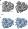
Figure 1
Figure 1. CryoEM reconstructions of the L. donovani and human ribosomes.
Stereo related views of the L. donovani (a) and human (b) ribosomes at 2.9 Å and 3.6 Å respectively. Approximate dimensions of the ribosome: 300 Å × 250 Å × 250 Å.
Figure 2
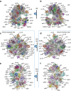
Figure 2. Arrangement of the L. donovani ribosome.
Atomic models of the whole ribosome (a,b), LSU (c,d), and SSU (e,f) of the L. donovani 80S ribosome. Protein molecules are shown in colour and rRNA molecules in grey. (c)The LSU as seen from the side exposed to solvent (Solvent exposed side). (d) The LSU as seen from the subunit interface (Subunit interface side). (e) The SSU as seen from the side exposed to solvent (Solvent exposed side). (f) The SSU as seen from the subunit interface (Subunit interface side). *Protein uL13 is involved in rRNA stabilization, binding the ends of srRNA2 and srRNA3.
Figure 3
Figure 3. Leishmanial ribosome expansion segments that are distinctive from non-trypanosomal ribosomes.
For clarity, the cryoEM map of the L. donovani ribosome was filtered to a resolution of 5 Å with the most conserved structure elements displayed in grey and the expansion segments distinctive from those in non-trypanosomal ribosomes in colour. (a,b) The solvent-exposed side (side visible from the exterior of the ribosome) of LSU (a) and SSU (b). (c,d) Whole ribosome with visible tRNA in the E site. CP, central protuberance.
Figure 4
Figure 4. Unique rRNA end localization in the (left) L. donovani ribosome compared with homologous regions in the (right) human ribosome.
Guide figures are rRNA components found in L. donovani and are related by two 90° turns, one along the x and one along the y axis. rRNA ends are denoted with green asterisks and labelled. The two 28S rRNA ends are labelled similarly for the human ribosome. For clarity, conserved 5S and 5.8S rRNA elements are shown in beige and are not labelled. Protein components are not shown. (a) Unique 3′ and 5′ ends of srRNA2 and srRNA3 compared with the homologous region in the human ribosome. (b) Unique 3′ and 5′ ends of srRNA4 compared with the homologous region in the human ribosome. (c) Unique 3′ end of LSU-β compared with the homologous region in the human ribosome. (d) The conserved 5′ end of LSU-α compared with the homologous region in the human ribosome. (e) Unique 5′ and 3′ ends of srRNA1, unique 3′ end of LSU- α and unique 5′ end of LSU-β compared with the homologous region in the human ribosome. Corresponding density and full models are shown in Supplementary Fig. 7.
Figure 5
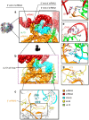
Figure 5. uL13-pin and rRNA fragmented end stabilization.
(a) Surface representation of the region surrounding uL13-pin (ribbon), which is the trypanosome-unique N-terminal extension of uL13, showing the locations of rRNA ends in relation to uL13-pin. KSD and eL14 are not shown for clarity. (b) Rotated view of the trapezoidal region in (a) showing the rRNA ring (surface) formed by LSU-β (cyan) and srRNA3 (orange) through which uL13-pin passes through. (c) Arrangement of the trypanosome-unique extension of eL33 which binds the 3′ end of srRNA3 (orange) adjacent to uL13. (d) Sandwiching of the 5′ end of srRNA3 (A3) between the 5′ end of srRNA2 (G1) and Arg14 of uL13. (e) Binding of the 3′ end of srRNA2 (C183) by Arg6 of uL13. (f) Interactions between srRNA3 and Arg11 of uL13. (g) Binding of the 3′ end of srRNA3 (A73) by eL33. Full models with density maps available in Supplementary Figs 10 and 11.
Figure 6
Figure 6. Functionally significant unique structural features in the L. donovani ribosome.
(a) The unusually large ES42L (blue) binds srRNA2 (red) and a C-terminal extension of eL28 (yellow and cyan) in L. donovani. eL28 rests in a groove which lies between two RNA helices in ES42L. The L. donovani unique portion of the extension is coloured cyan, superimposed against that in T. brucei (magenta). (b,c) Boxed regions of (a) showing interactions between the C-terminal extension of eL28 and ES42L. (d) A closeup, 180° rotated view of the boxed region in (a), showing the kissing helix interaction between srRNA2 and ES42L formed by canonical basepairing. (e) Positioning of eS17 in L. donovani and T. brucei in relation to the trypanosome unique portions of ES6S and ES7S. Dashed line depicts flexible region of ES7S visible in the cryoEM map.
Figure 7
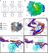
Figure 7. Human ribosome structure and insights into paromomycin specificity.
(a) Decoding A-site sequences of Homo sapiens, L. donovani, and Thermus thermophilus ribosomes. The binding site of paromomycin is boxed in this sequence representation. Red text denotes the 1,409–1,491 basepair which is broken in H. sapiens and other eukaryotes but restored in L. donovani. (b) CryoEM reconstruction of the human 80S ribosome at 3.6 Å coloured by radius. (c) Superimposition of the decoding A-sites of the human and the L. donovani ribosomes with that of the T. thermophilus ribosome complexed with paromomycin. Bases of 1,492 and 1,493 are hidden for clarity. (d) Comparison of the 1,409–1,491 basepair displayed in (c) in H. sapiens, L. donovani and T. thermophilus ribosomes, highlighting the unfavourable bonding angle between 1,409 and 1,491 (C1710 and A1823 human numbering) in the human ribosome (blue). Paromomycin displayed as a space-filling model to illustrate the severe clash with A1823 of the human decoding site. (e) A 20° related view of (d) along the x-axis. Black text indicates E. coli numbering, and coloured text corresponds to the species colour legend (orange, L. donovani; dark purple, T. thermophilus; blue, H. sapiens).

Similar articles

Cited by

References

    1. World Health Organization. Control of the leishmaniases. World Health Organization Tech Rep Ser 949, 67–71 (2010). - PubMed
    1. Anger A. M. et al.. Structures of the human and Drosophila 80S ribosome. Nature 497, 80–85 (2013). - PubMed
    1. Hashem Y. et al.. High-resolution cryo-electron microscopy structure of the Trypanosoma brucei ribosome. Nature 494, 385–389 (2013). - PMC - PubMed
    1. Khatter H., Myasnikov A. G., Natchiar S. K. & Klaholz B. P. Structure of the human 80S ribosome. Nature 520, 640–645 (2015). - PubMed
    1. Behrmann E. et al.. Structural snapshots of actively translating human ribosomes. Cell 161, 845–857 (2015). - PMC - PubMed

Publication types

MeSH terms