. 2018 Feb;23(2):467-475.
doi: 10.1038/mp.2016.184. Epub 2016 Oct 18.

DIXDC1 contributes to psychiatric susceptibility by regulating dendritic spine and glutamatergic synapse density via GSK3 and Wnt/β-catenin signaling

Affiliations

DIXDC1 contributes to psychiatric susceptibility by regulating dendritic spine and glutamatergic synapse density via GSK3 and Wnt/β-catenin signaling

P-M Martin et al. Mol Psychiatry. 2018 Feb.

Abstract

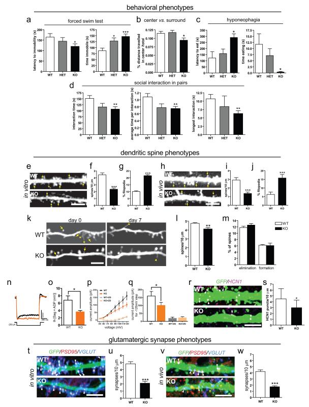
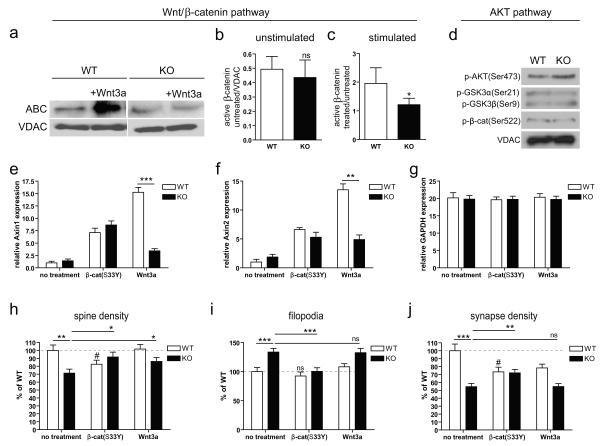
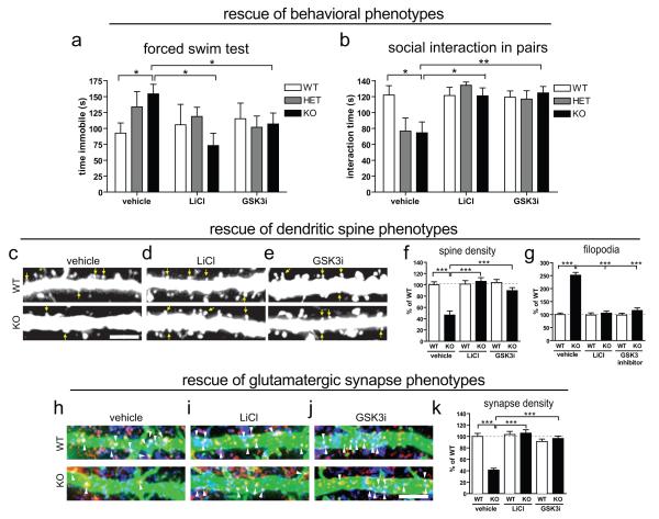
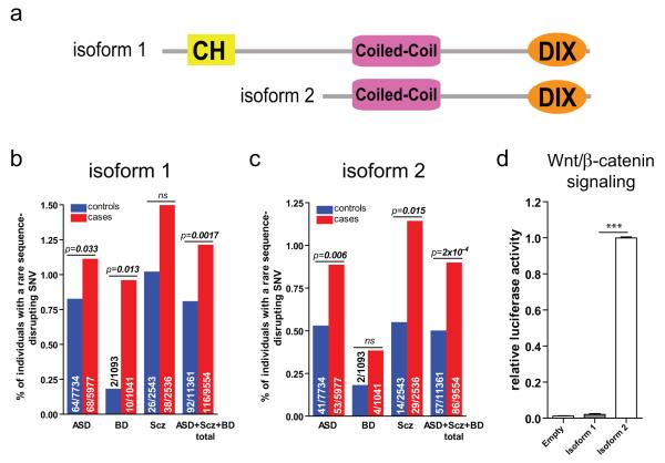
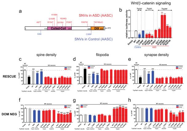
Mice lacking DIX domain containing-1 (DIXDC1), an intracellular Wnt/β-catenin signal pathway protein, have abnormal measures of anxiety, depression and social behavior. Pyramidal neurons in these animals' brains have reduced dendritic spines and glutamatergic synapses. Treatment with lithium or a glycogen synthase kinase-3 (GSK3) inhibitor corrects behavioral and neurodevelopmental phenotypes in these animals. Analysis of DIXDC1 in over 9000 cases of autism, bipolar disorder and schizophrenia reveals higher rates of rare inherited sequence-disrupting single-nucleotide variants (SNVs) in these individuals compared with psychiatrically unaffected controls. Many of these SNVs alter Wnt/β-catenin signaling activity of the neurally predominant DIXDC1 isoform; a subset that hyperactivate this pathway cause dominant neurodevelopmental effects. We propose that rare missense SNVs in DIXDC1 contribute to psychiatric pathogenesis by reducing spine and glutamatergic synapse density downstream of GSK3 in the Wnt/β-catenin pathway.


Figures

Figure 1
Figure 1
Phenotypes in Dixdc1KO mice. (a-d) Behavioral phenotypes. (a) Despair (FST). (b) Anxiety (time in center vs. surround in an open field). (c) Drive to eat vs. anxiety over unfamiliar (Hyponeophagia assay). (d) Social behavior (SIP). (e-s) Dendritic spine phenotypes: Primary dendrites of cultured neurons transfected with GFP (e quantified in f and g). Top, WT; bottom, KO; arrow, mature spine; asterisk, filopodial projection. Cortical L5/6 apical dendrites in brain tissue labeled by Tg(Thy1-EGFP)M (h quantified in i and j). Top, WT; bottom, KO; arrows and asterisks as in (e). (k-m) Imaging of spine density and spine dynamics in vivo. 2-photon images of the same dendritic branch over 7 days labeled by Tg(Thy1-YFP)H in the primary somatosensory cortex of living adult mice (k quantified in l and m). Top, WT; bottom, KO; arrow, eliminated spine; arrowhead, newly-formed spine. (n-q) Recording of Ih-dependent voltage and current. (n) Representative whole cell current clamp recordings in L5/6 prefrontal corticothalamic neurons; black, WT; orange, KO. (o) Quantification of voltage sag and rebound (ADP) dependent of Ih. (p) Direct measurement of hyperpolarization-activated currents and inhibition by ZD7288, a specific antagonist of Ih; black, WT; orange, KO; grey, ZD7288 treated WT; light orange, ZD7288 treated KO. (q) Ih current activation at −120mV potential step; colors as in p. (r-s) Immunohistological visualization of HCN1 channel protein: Representative micrographs of cortical L5/6 apical dendrites (r quantified in s). Top, WT; bottom, KO; far red, HCN1; green (Tg(Thy1-EGFP)M). (t-w) Glutamatergic synapse phenotypes. Co-localized immunohistochemical markers for glutamatergic synapses on dendrites in GFP-transfected cultured neurons (t quantified in u), and cortical L5/6 apical dendrites labeled by Tg(Thy1-EGFP)M (v quantified in w). Top, WT; bottom, KO; blue, VGLUT; red, PSD95; green, GFP; arrowheads, colocalized green/blue/red puncta. Scale bars, 5 μm. *p≤0.05; **p≤0.01; ***p≤0.001
Figure 2
Figure 2
Decreased Wnt/β-catenin signal transduction in Dixdc1KO neurons. (a) Immunoblot of active β-catenin (ABC) from cultured neurons; (b-c) Quantification in unstimulated (b) and Wnt3a-stimulated (c) WT vs. KO neurons. (d) AKT pathway: Immunoblots of phosphorylated AKT (p-AKT(Ser473)), AKT-phosphorylated GSK3 (p-GSK3α(Ser21) top, p-GSK3β(Ser9) bottom, and AKT-phosphorylated β-catenin (p-β-cat(Ser522)), in WT vs. KO neurons. (e-g) Q-PCR for Wnt/β-catenin pathway transcriptional targets in neurons: Axin1 (e) and Axin2 (f); GAPDH (control) (g); KO (black) vs. WT (white). Left, untreated; Middle, transfected with β-cat(S33Y); Right, treated with Wnt3a. (h-j) Neurodevelopmental responses to Wnt/β-catenin pathway stimulation. Quantification (as in Fig 1): (h) spine density, (i) % filopodia and (j) glutamatergic synapse density; white, WT; black, KO, all conditions normalized to WT untreated (left-most bar). Comparisons as in (e-g). *p≤0.05; **p≤0.01; ***p≤0.001; #p≤0.05 vs. untransfected WT.
Figure 3
Figure 3
GSK3 inhibition rescues Dixdc1KO phenotypes. (a-b) Pharmacologic rescue of behavioral phenotypes. The FST phenotype (increased time immobile) (a) and SIP phenotype (reduced social interaction) (b) are rescued by injection of either lithium (middle) or GSK3i (right). (c-g) Pharmacologic rescue of dendritic spine phenotypes. Cortical L5/6 apical dendrites labeled by Tg(Thy1-EGFP)M in mice treated with vehicle (c), lithium (d) or GSK3i (e). Top, WT; bottom, KO; arrows and asterisks as in Fig 1e. Quantification, all conditions normalized to WT vehicle (left-most bar) (f and g). (h-k) Pharmacologic rescue of glutamatergic synapse phenotypes. Co-localized immunohistochemical markers for glutamatergic synapses on cortical L5/6 apical dendrites labeled by Tg(Thy1-EGFP)M in mice treated with vehicle (h), lithium (i) or GSK3i (j). Top, WT; bottom, KO; colors as in Fig 1r. Quantification with all conditions normalized to WT vehicle (left-most bar) (k). Scale bars, 5μm. *p≤0.05; **p≤0.01; ***p≤0.001
Figure 4
Figure 4
Increased rare sequence-disrupting DIXDC1 SNVs in ASD, BD and Scz. (a) Schematic representation of human DIXDC1 isoform 1 and isoform 2. Yellow, calponin homology domain (CH); pink, coiled-coil domain; orange, DIX domain. (b, c) % of individuals carrying a rare sequence-disrupting (nonsense, missense or splice-site disrupting) SNV in DIXDC1 isoform 1 (b) and 2 (c). (d) Relative TOPflash (Wnt/β-catenin signaling) activity for WT human DIXDC1 isoform 1 vs. isoform 2; values normalized to isoform 2 (white bar). ***p≤0.001
Figure 5
Figure 5
Rescue and dominant neurodevelopmental effects of missense SNVs with functional effects on the Wnt/β-catenin pathway. (a) Schematic diagram of isoform 2 indicating positions of rare missense SNVs found in ASD (red, top) vs. ethnically matched-controls (blue, bottom) in the discovery (AASC) dataset. (b) TOPflash for alleles shown in (a); values normalized to WT isoform 2 (white bar); alleles grouped as hypoactive, neutral, or hyperactive based on Δ ≥ 10% vs. WT. (c-e) Neurodevelopmental rescue: WT human DIXDC1 isoform 2 (grey bar) restores spine density (c), % immature spines (d) and glutamatergic synapse density (e) to WT levels (white bar) when expressed in KO neurons. Control-derived SNVs (K89I, C389F) (blue bars) but no case-derived SNVs (red bars), similarly rescue these KO phenotypes. (f-h) Dominant neurodevelopmental effects: WT human DIXDC1 isoform 2 (grey bar) or cDNAs containing hypoactive or activity-neutral SNVs (K89I, C389F, R154T, R249Q, T401M, P285T) do not cause neurodevelopmental phenotypes, but cDNAs containing hyperactive SNVs found in cases (A87T, Q169R, K188N, Q218H, R367W) decrease spine density (f), increase immature spine % (g) and decrease glutamatergic synapse density (h) when overexpressed in WT neurons. *p≤0.05; **p≤0.01; ***p≤0.001 vs. WT in (b-e); ##p≤0.01; ###p≤0.001 vs. identical genotype + empty vector in (c-h).

Similar articles

Cited by

References

    1. Pinto D, Delaby E, Merico D, Barbosa M, Merikangas A, Klei L, et al. Convergence of genes and cellular pathways dysregulated in autism spectrum disorders. Am J Hum Genet. 2014 May 1;94(5):677–94. - PMC - PubMed
    1. De Rubeis S, He X, Goldberg AP, Poultney CS, Samocha KE, Cicek AE, et al. Synaptic, transcriptional and chromatin genes disrupted in autism. Nature. 2014 - PMC - PubMed
    1. Mulligan KA, Cheyette BN. Wnt signaling in vertebrate neural development and function. J Neuroimmune Pharmacol. 2012 Dec;7(4):774–87. - PMC - PubMed
    1. Krumm N, O'Roak BJ, Shendure J, Eichler EE. A de novo convergence of autism genetics and molecular neuroscience. Trends Neurosci. 2014 Feb;37(2):95–105. - PMC - PubMed
    1. Shiomi K, Uchida H, Keino-Masu K, Masu M. Ccd1, a novel protein with a DIX domain, is a positive regulator in the Wnt signaling during zebrafish neural patterning. Curr Biol. 2003 Jan 8;13(1):73–7. - PubMed

Publication types

MeSH terms

Substances