Temporomandibular Joint Disc Repositioning Using an Orthopedic Suture Anchor: A Modified Disc Anchoring Technique
- PMID: 27752215
- PMCID: PMC5048315
- DOI: 10.1007/s12663-015-0838-6
Temporomandibular Joint Disc Repositioning Using an Orthopedic Suture Anchor: A Modified Disc Anchoring Technique
Abstract
Purpose: The study assessed the efficacy of orthopedic suture anchor as a modified suture anchor for disc repositioning in case of a closed lock of TMJ.
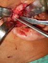
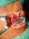
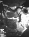
Patient and methods: Disc repositioning was undertaken via a mini preauricular approach. The disc was repositioned on the surface of the condyle and stabilized using an orthopedic suture anchor. Postoperatively functional outcomes were assessed in terms of reduction in pain, joint movement and absence of joint noise and clicking sounds. Postoperative MRI was used to assess the disc position and morphological changes in the disc and arthritic changes in the condyle at the end of six months.
Results: Patients were post surgically followed up at regular intervals of 1, 3 and 6 months. Patient experienced significant improvement in mouth opening with good functional outcomes and stable repositioning of disc as noticed By MRI at the end of six months.
Conclusions: We describe a modified technique of disc repositioning using an orthopedic suture anchor for more favorable disc position and joint function. However the long term functional sequel of the procedure and changes in the articular disc needs to be assessed.
Keywords: Disc repositioning; Internal derangement; Orthopedic suture anchor.
Figures


Similar articles
-
Clinical and MRI Evaluation of Orthodontic Mini-Screws for Disc Repositioning in Internal Derangement of TMJ: A Prospective Study.J Maxillofac Oral Surg. 2018 Mar;17(1):52-58. doi: 10.1007/s12663-017-1018-7. Epub 2017 May 15. J Maxillofac Oral Surg. 2018. PMID: 29382994 Free PMC article.
-
The Mitek mini anchor for TMJ disc repositioning: surgical technique and results.Int J Oral Maxillofac Surg. 2001 Dec;30(6):497-503. doi: 10.1054/ijom.2001.0163. Int J Oral Maxillofac Surg. 2001. PMID: 11829231
-
Modified Temporomandibular Joint Disc Repositioning With Mini-screw Anchor: Part II-Stability Evaluation by Magnetic Resonance Imaging.J Oral Maxillofac Surg. 2019 Feb;77(2):273-279. doi: 10.1016/j.joms.2018.07.016. Epub 2018 Jul 24. J Oral Maxillofac Surg. 2019. PMID: 30118666
-
Disc repositioning: does it really work?Oral Maxillofac Surg Clin North Am. 2015 Feb;27(1):85-107. doi: 10.1016/j.coms.2014.09.007. Oral Maxillofac Surg Clin North Am. 2015. PMID: 25483446 Review.
-
Role of magnetic resonance imaging in the clinical diagnosis of the temporomandibular joint.Cells Tissues Organs. 2005;180(1):6-21. doi: 10.1159/000086194. Cells Tissues Organs. 2005. PMID: 16088129 Review.
References
-
- Cai XY, Jin JM, Yang C. Changes in disc position, disc length, and condylar height in the temporomandibular joint with anterior disc displacement: a longitudinal retrospective magnetic resonance imaging study. J Oral Maxillofac Surg. 2011;69(11):e340–e346. doi: 10.1016/j.joms.2011.02.038. - DOI - PubMed
-
- Quinn PD (1998) Color atlas of temporomandibular joint surgery. In: Louis St (ed) Surgery for internal derangements, chap 4. Mosby, pp 55–99
LinkOut - more resources
Full Text Sources
Other Literature Sources
