. 2016 Oct 12:10:12.
doi: 10.1186/s13036-016-0033-4. eCollection 2016.

Design and engineering of a transmissible antiviral defense

Affiliations

Design and engineering of a transmissible antiviral defense

Matthew L Paff et al. J Biol Eng. .

Abstract

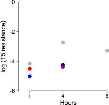
Background: We propose, model, and implement a novel system of population-level intervention against a virus. One context is a treatment against a chronic infection such as HIV. The underlying principle is a form of virus 'wars' in which a benign, transmissible agent is engineered to protect against infection by and spread of a lethal virus. In our specific case, the protective agent consists of two entities, a benign virus and a gene therapy vector mobilized by the benign virus.

Results: Numerical analysis of a mathematical model identified parameter ranges in which adequate, population-wide protection is achieved. The protective system was implemented and tested using E. coli, bacteriophage M13 and a phagemid vector mobilized by M13 to block infection by the lethal phage T5. Engineering of M13 profoundly improved its dynamical properties for facilitating spread of the gene therapy vector. However, the gene therapy vector converts the host cell to resist T5 too slowly for protection on a time scale appropriate for T5.

Conclusions: Overall, there is a reasonable marriage between the mathematical model and the empirical system, suggesting that such models can be useful guides to the design of such systems even before the models incorporate most of the relevant biological details.

Keywords: Bacteriophage; Genetic engineering; Transmissible vaccine; Virus.


Figures

Fig. 1
Fig. 1
Protection against T5 infection provided by the phagemid-encoded Targetron. Cells were infected by the ampicillin-resistant phagemid (vaccine) at an MOI of 1.0 (phagemid stock also contained a low concentration of helper phage at MOI 0.01) and grown for the time indicated. They were plated separately on ampicillin and on a high density of T5. The log10 proportion of ampicillin-resistant cells that are T5-resistant is shown. Resistance levels are never more than 1 % and often not more than 0.1 %, even after 8 h. Colors indicate replicates; only one sample was assayed at 8 h
Fig. 2
Fig. 2
Effect of initial vaccinations and reproductive parameters on dynamics of vaccine and helper. All bacteria ultimately become infected with helper, but protection by vaccine accrues only to the bacteria that (also) receive vaccine. Furthermore, vaccine can only be acquired before a cell is infected with helper, so cells infected with helper-only can never become protected. Thus we care most about the long term fraction of all cells infected with vaccine regardless of whether they are also infected with helper (thick dark red). a Using parameters from Table 4 in Appendix, and introducing only 100 initial vaccinations (as doubly-infected cells at time 0) the vaccine is vastly outpaced by helper by nearly 4 logs and is ineffective at protecting the population. Progressively higher numbers of initial vaccinations yield progressively better vaccine coverage [(b), (c)]. Panel d increases the reproductive output 10-fold for doubly-infected cells (b VM=7, b MV=0.06) but otherwise uses the same conditions as in (b): there is a substantial improvement in coverage. Both c and d result in most infected cells carrying vaccine. Thus improvement in reproductive parameters and high levels of ‘manual’ vaccination are different ways to substantially increase coverage. Equations used are given in (A1) with lethal virus omitted; parameter values were as in Table 4 in Appendix, except as indicated for (D). Initial vaccinations were added as H VMS at time 0
Fig. 3
Fig. 3
Vaccine success rates depend on manual vaccinations and the reproductive properties of vaccine and helper. Each panel shows the results from hundreds of numerical simulations stopped at time 400. For each combination of b M and b MV tested, the proportion of bacterial hosts with vaccine over all infected bacteria (at time 400) is indicated with color – purple is best, yellow worst. Color opacity is scaled in proportion to the fraction of infected bacteria over all bacteria. As most simulations resulted in all or nearly all bacteria becoming infected with either vaccine or helper by time 400, most of the panel is solid color. The white space in the lower left region is highly transparent because those parameter values resulted in few bacteria infected; zones with intermediate levels of infection are narrow. Each panel assumes a fixed ratio of vaccine/helper output (b VM:b MV) and a specific number of initial vaccinations (100 or 100,000), as indicated. Black circles indicate the approximate behavior of wildtype M13 and phagemid (a, c) and of engineered M13 and phagemid (b, d) and are placed on the panels most closely representing the empirical relationship of b VM: b MV. The benefit of engineering a vaccine with a large reproductive excess over helper is readily evident, as is the value of introducing larger numbers of vaccinations. Equations were those of (A1), with lethal virus omitted and all forms of vaccine-infected cells combined; parameter values were those of Table 4 in Appendix, except for b M, b MV and b VM, which were varied and are given in the figure. The initial density of uninfected cells was 108; vaccinated cells were introduced as H VMS at time 0
Fig. 4
Fig. 4
Numerical dynamics of lethal virus, host and two-component vaccine. Manual vaccinations were introduced as doubly-infected cells (H VMS) at time 0. In the long term, the population comes to consist of only doubly-infected cells (H VMR, thick dark red) and lethal virus (L, purple), but the early dynamics depend on initial conditions and the rate at which vaccine-infected cells convert from sensitive to resistance against the lethal virus (given by c). a b c: The cases of c=0.1, c=0.01 and c=0.001 are contrasted and show a large effect of delayed protection. With c=0.001, many cells carrying vaccine are killed because they were not yet converted to a resistant state when exposed to lethal virus. d The rates of vaccine and helper production from doubly-infected cells are increased 10-fold over those in (a), but parameters and initial conditions are otherwise the same. The result is a nearly 1-log increase in protected cells in the time frame shown. e f: Burst size of the lethal virus is dropped 10-fold over that in (a). Parameters and initial conditions are otherwise the same as in (a) for panel (e), but the number of initial vaccinations (H VMS) is increased 10-fold in (f), resulting in a 7-fold increase in protected cells. In these latter two trials, the lethal virus has effectively no effect on the bacterial dynamics in the time frame shown. For all panels, equations used are given in (A1), parameters as in Table 4 in Appendix. Unless indicated otherwise, runs started with 108 uninfected cells, 105 doubly-infected (sensitive), 1000 lethal virus, and a carrying capacity of 109
Fig. 5
Fig. 5
Empirical dynamics of the two component vaccine in absence of the lethal virus. Three replicates are shown (a)–(c), conducted at different times but all attempting to repeat the same initial conditions. All replicates have several properties in common. (i) There is an increase in cells infected with helper-only from undetectable at time 0 to an abundance exceeding all other types of infections at 3hr (the limit of detection is the dashed line at 103). (ii) Cells infected with vaccine-only (pgT) increase most substantially in the first hour, then about 10-fold in the next 2 h; much of the latter could be explained by cell growth, but the first-hour growth must be from infection. (iii) Doubly-infected cells increase slowly in the first hour but 100-fold in the next 2 h for two of the trials. A 100-fold increase is too high to be explained by cell growth and thus must be partly from infection. There are also obvious differences among replicates. (iv) The doubly-infected cell density at 3 h is usually around 107 despite a 10-fold variation in the inoculum. Open symbols, which lie at the limit of detection, indicate that no bacteria were detected. Colored dashed lines indicate that the true slope is undetermined because at least the early time point is unknown
Fig. 6
Fig. 6
Dynamics with both phages. a, b Two replicates in which phage T5 was added to a culture of E. coli that had been infected and grown overnight with vaccine/phagemid and helper (dashed lines) or added to a culture that was not exposed to phagemid or helper (solid lines). (Vaccine and helper were added at a multiplicity of infection of 1.0 and 0.01, respectively, at initiation of the overnight culture). There is a sharp rise in T5 density after addition regardless of the presence of the vaccine (blue curves). However, cell density rebounds in the vaccine-protected population from growth of the survivors, which are resistant. Due to the effect of helper on cell growth, a higher initial density of cells is present in the unprotected culture than in the protected culture. c Drop in host density compared between presence and absence of the vaccine. The decline in cell density from 2hr to 4h is only 0.75 and 1.15 logs less when the vaccine is present than when it is absent (comparing a and b)

Similar articles

References

    1. Richman DD. Antiviral Drug Resistance. Chichester; New York: Wiley; 1996.
    1. Barrett AD, Dimmock NJ. Defective interfering viruses and infections of animals. Curr Top Microbiol Immunol. 1986;128:55–84. doi: 10.1007/978-3-642-71272-2_2. - DOI - PubMed
    1. Marriott AC, Dimmock NJ. Defective interfering viruses and their potential as antiviral agents. Rev Med Virol. 2010;20(1):51–62. doi: 10.1002/rmv.641. - DOI - PubMed
    1. Mebatsion T, Finke S, Weiland F, Conzelmann KK. A CXCR4/CD4 pseudotype rhabdovirus that selectively infects HIV-1 envelope protein-expressing cells. Cell. 1997;90(5):841–7. doi: 10.1016/S0092-8674(00)80349-9. - DOI - PubMed
    1. Schnell MJ, Johnson JE, Buonocore L, Rose JK. Construction of a novel virus that targets HIV-1-infected cells and controls HIV-1 infection. Cell. 1997;90(5):849–57. doi: 10.1016/S0092-8674(00)80350-5. - DOI - PubMed

LinkOut - more resources