. 2016 Oct 6;3(5):ENEURO.0274-16.2016.
doi: 10.1523/ENEURO.0274-16.2016. eCollection 2016 Sep-Oct.

Terminal Dopamine Release Kinetics in the Accumbens Core and Shell Are Distinctly Altered after Withdrawal from Cocaine Self-Administration

Affiliations

Terminal Dopamine Release Kinetics in the Accumbens Core and Shell Are Distinctly Altered after Withdrawal from Cocaine Self-Administration

Michael P Saddoris. eNeuro. .

Abstract

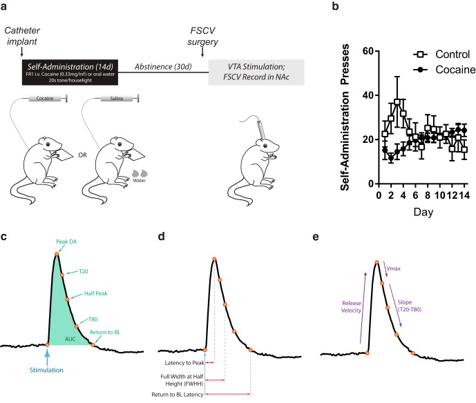
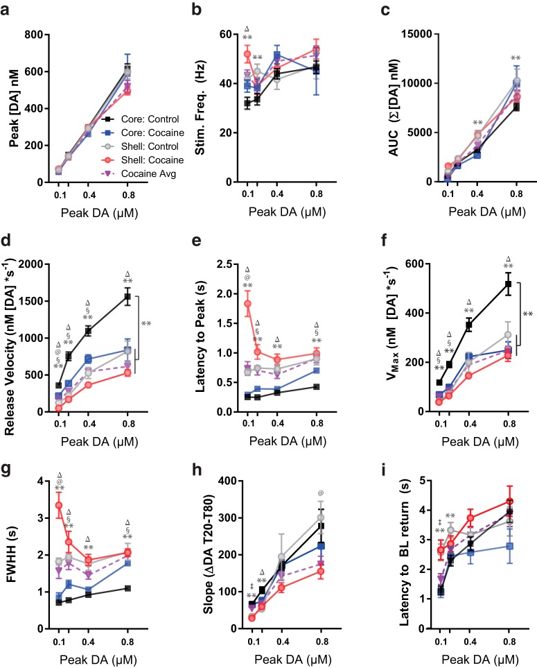
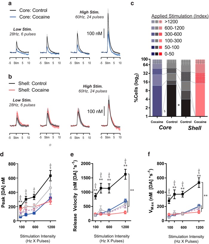
Repeated self-administration of cocaine is associated with impairments in motivated behaviors as well as alterations in both dopamine (DA) release and neural signaling within the nucleus accumbens (NAc). These impairments are present even after several weeks of abstinence from drug taking, suggesting that the self-administration experience induces long-lasting neuroplastic alterations in the mesolimbic DA circuit. To understand these changes at the terminal level, rats were allowed to self-administer either cocaine intravenously (∼1 mg/kg per infusion) or water to a receptacle (control) in 2-h sessions over 14 days, followed by 30 days of enforced abstinence. Fast-scan cyclic voltammetry was used to record real-time DA release in either NAc core or shell after electrical stimulations of the ventral tegmental area (VTA) in freely-moving animals. In controls, the kinetics of DA release in the core and shell strikingly differed, with shell displaying slower release and reuptake rates than core. However, cocaine experience differentially altered these signaling patterns by NAc subregion. In the shell, cocaine rats showed less sensitivity to the dynamic range of applied stimulations than controls. In the core, by contrast, cocaine rats displayed robustly reduced peak DA release given the same stimulation, while also showing slower release and reuptake kinetics. The differential effects of cocaine self-administration on terminal function between core and shell is consistent with a region-specific functional reorganization of the mesolimbic DA system after repeated exposure and may provide an anatomical substrate for altered cognitive function after chronic drug-taking and addiction.

Keywords: Michaelis–Menten; dopamine transporter; drug addiction; plasticity; ventral striatum; voltammetry.


Figures

Figure 1.
Figure 1.
Schematic of experimental design, reinforced presses across the 14 d of self-administration training, and schematic of different metrics of DA release kinetics. A, Points in the release kinetics in relation to the peak DA release (i.e., point of greatest [DA] after stimulation). Half peak is the point at exactly half of peak concentration, Return to baseline (BL) is the point at which the [DA] was within a 95% confidence interval of the baseline, and T20 and T80 reflect 20% and 80% decrease in [DA] from peak, respectively. AUC was estimated by summing the [DA] in each 100-ms bin between stimulation and return to BL. B, Latency measures derived from the points of release and reuptake from A. Latency to peak, FWHH (i.e., latency from stimulation to half peak), and return to BL latency are relative to stimulation, whereas T20 and T80 latencies are relative to peak. C–E, Rates of change relative to points during release. Release velocity is the rate of increase in [DA] from stimulation to peak, Vmax is the rate of uptake between the peak and T20, and slope is the rate of uptake between T20 and T80.
Figure 2.
Figure 2.
Placement of electrodes during recording in controls (top) or cocaine (bottom) rats. Black circles, core; gray circles, shell.
Figure 3.
Figure 3.
Distribution of peak [DA] amplitude from stimulation trials in the NAc core (control, black; cocaine, blue) and NAc shell (control, gray; cocaine, red). Peak [DA] responses for each stimulation were binned by 50-ms epochs from 0 to 1200 nm, while all stimulations that were greater than 1200 nm represented the final bin. Proportion reflected the number of stimulations in that bin as a proportion of all stimulations from that group. **Control core vs. control shell; §control core vs. cocaine core; @control shell vs. cocaine shell; p < 0.001 for relevant χ2.
Figure 4.
Figure 4.
Representative color plots of stimulated DA release in NAc core (A and B) and NAc shell (C and D). E, Overlapped traces of DA elicited by electrical stimulation in core and shell of controls and cocaine-experienced subjects from the representative color plots in A–D.
Figure 5.
Figure 5.
Kinetic factors of DA release aligned by peak [DA] in control core (black squares), cocaine core (blue squares), control shell (gray circles), and cocaine shell (red circles) recordings. **Control core vs. control shell; Δcontrol core vs. both cocaines; §control core vs. cocaine core; @control shell vs. cocaine shell; control Shell vs. both cocaines; p < 0.01 (Bonferroni-corrected α for multiple comparisons).
Figure 6.
Figure 6.
Average phasic DA release in the NAc core (A) and shell (B) of controls (black/gray) and cocaine self-administering rats (blue/red) in stimulation index–aligned bins. C, For each drug group and region, the proportion of cells (of all observations) in each stimulation index bin. Note log2 scale used to show the loss specifically of the low stimulation index observations in the cocaine groups. Peak [DA] (D), rise velocity (E), and Vmax (F) for treatment groups across stimulation intensity bins. **Control core vs. control shell; Δcontrol core vs. both cocaines; §control core vs. cocaine core; @control shell vs. cocaine shell; control shell vs. both cocaines; p < 0.01 (Bonferroni-corrected α for multiple comparisons).

Similar articles

Cited by

References

    1. Addy NA, Daberkow DP, Ford JN, Garris PA, Wightman RM (2010) Sensitization of rapid dopamine signaling in the nucleus accumbens core and shell after repeated cocaine in rats. J Neurophysiol 104:922–931. 10.1152/jn.00413.2010. 10.1152/jn.00413.2010 - DOI - PMC - PubMed
    1. Aragona BJ, Cleaveland NA, Stuber GD, Day JJ, Carelli RM, Wightman RM (2008) Preferential enhancement of dopamine transmission within the nucleus accumbens shell by cocaine is attributable to a direct increase in phasic dopamine release events. J Neurosci 28:8821–8831. 10.1523/JNEUROSCI.2225-08.2008 - DOI - PMC - PubMed
    1. Belin D, Everitt BJ (2008) Cocaine seeking habits depend upon dopamine-dependent serial connectivity linking the ventral with the dorsal striatum. Neuron 57:432–441. 10.1016/j.neuron.2007.12.019 - DOI - PubMed
    1. Belin D, Jonkman S, Dickinson A, Robbins TW, Everitt BJ (2009) Parallel and interactive learning processes within the basal ganglia: relevance for the understanding of addiction. Behav Brain Res 199:89–102. 10.1016/j.bbr.2008.09.027 - DOI - PubMed
    1. Berridge KC (2012) From prediction error to incentive salience: mesolimbic computation of reward motivation. European J Neurosci 35:1124–1143. 10.1111/j.1460-9568.2012.07990.x - DOI - PMC - PubMed

Publication types

MeSH terms

LinkOut - more resources