

Reduced Protein Expression of the Na+/Ca2++K+-Exchanger (SLC24A4) in Apical Plasma Membranes of Maturation Ameloblasts of Fluorotic Mice
- PMID: 27752731
- PMCID: PMC5215084
- DOI: 10.1007/s00223-016-0197-4
Reduced Protein Expression of the Na+/Ca2++K+-Exchanger (SLC24A4) in Apical Plasma Membranes of Maturation Ameloblasts of Fluorotic Mice
Abstract
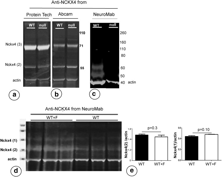
Exposure of forming enamel to fluoride results into formation of hypomineralized enamel. We tested whether enamel hypomineralization was caused by lower expression of the NCKX4/SLC24A4 Ca2+-transporter by ameloblasts. Three commercial antibodies against NCKX4 were tested on enamel organs of wild-type and Nckx4-null mice, one of which (a mouse monoclonal) was specific. This antibody gave a prominent staining of the apical plasma membranes of maturation ameloblasts, starting at early maturation. The layer of immuno-positive ameloblasts contained narrow gaps without immunostaining or with reduced staining. In fluorotic mouse incisors, the quantity of NCKX4 protein in ameloblasts as assessed by western blotting was not different from that in non-fluorotic ameloblasts. However, immunostaining of the apical plasma membranes of fluorotic ameloblasts was strongly reduced or absent suggesting that trafficking of NCKX4 to the apical membrane was strongly reduced. Exposure to fluoride may reduce NCKX4-mediated transport of Ca2+ by maturation stage ameloblasts which delays ameloblast modulation and reduces enamel mineralization.
Keywords: Calcium; Enamel fluorosis; Mechanism; Null mutation; Transport.
Conflict of interest statement
A. L. J. J. Bronckers, R. Jalali, J. Lytton declare that they have no conflict of interest. Human and Animal Rights and Informed Consent The authors confirm that the animal studies were approved and carried out according to national guidelines.
Figures
Similar articles
-
Ameloblast Modulation and Transport of Cl⁻, Na⁺, and K⁺ during Amelogenesis.J Dent Res. 2015 Dec;94(12):1740-7. doi: 10.1177/0022034515606900. Epub 2015 Sep 24. J Dent Res. 2015. PMID: 26403673 Free PMC article.
-
The anion exchanger Ae2 is required for enamel maturation in mouse teeth.Matrix Biol. 2008 Mar;27(2):119-27. doi: 10.1016/j.matbio.2007.09.006. Epub 2007 Oct 11. Matrix Biol. 2008. PMID: 18042363 Free PMC article.
-
Expression of the sodium/calcium/potassium exchanger, NCKX4, in ameloblasts.Cells Tissues Organs. 2012;196(6):501-9. doi: 10.1159/000337493. Epub 2012 Jun 5. Cells Tissues Organs. 2012. PMID: 22677781 Free PMC article.
-
Ion Transport by Ameloblasts during Amelogenesis.J Dent Res. 2017 Mar;96(3):243-253. doi: 10.1177/0022034516681768. Epub 2016 Dec 19. J Dent Res. 2017. PMID: 28221098 Review.
-
Cellular and chemical events during enamel maturation.Crit Rev Oral Biol Med. 1998;9(2):128-61. doi: 10.1177/10454411980090020101. Crit Rev Oral Biol Med. 1998. PMID: 9603233 Review.
Cited by
-
Calmodulin binds and modulates K+-dependent Na+/Ca2+-exchanger isoform 4, NCKX4.J Biol Chem. 2021 Jan-Jun;296:100092. doi: 10.1074/jbc.RA120.015037. Epub 2020 Nov 23. J Biol Chem. 2021. PMID: 33199372 Free PMC article.
-
Correlations Between Single Nucleotide Polymorphisms, Cognitive Dysfunction, and Postmortem Brain Pathology in Alzheimer's Disease Among Han Chinese.Neurosci Bull. 2019 Apr;35(2):193-204. doi: 10.1007/s12264-019-00343-2. Epub 2019 Feb 19. Neurosci Bull. 2019. PMID: 30783964 Free PMC article.
-
Expression of potassium-dependent sodium-calcium exchanger in the murine lens.Exp Eye Res. 2018 Feb;167:18-24. doi: 10.1016/j.exer.2017.11.002. Epub 2017 Nov 9. Exp Eye Res. 2018. PMID: 29128492 Free PMC article.
-
The Na+/Ca2+, K+ exchanger NCKX4 is required for efficient cone-mediated vision.Elife. 2017 Jun 26;6:e24550. doi: 10.7554/eLife.24550. Elife. 2017. PMID: 28650316 Free PMC article.
-
Regulation of K+-Dependent Na+/Ca2+-Exchangers (NCKX).Int J Mol Sci. 2022 Dec 29;24(1):598. doi: 10.3390/ijms24010598. Int J Mol Sci. 2022. PMID: 36614039 Free PMC article. Review.
References
MeSH terms
Substances
LinkOut - more resources
Full Text Sources
Other Literature Sources
Molecular Biology Databases
Miscellaneous
